Squamous epithelium in the cytology of the cervix. Liquid cytology method: we diagnose the presence or absence of cervical cancer without errors. How is liquid cytology performed?
The main problem of oncology in general is the diagnosis of the tumor process at the earliest stage and the timely treatment of malignant neoplasms. For this, modern diagnostic methods are used, which make it possible to determine the presence of atypical cells in any tissue of the body.
Oncogynecology is an important part of preserving the reproductive health of the female population. Diagnosis of malignant neoplasms of the genital organs is the main task of this section of medicine. Cytological examination of scrapings of the cervix and cervical canal is an integral part of the diagnosis of genital tumors in women. But not all women know:
- which specialist prescribes the study;
- why and how it is carried out;
- what results can be obtained;
- how to interpret them.
Women who have been assigned such an examination are also concerned about other issues. The answers to them can be obtained by reading this article.
What is a cytological examination of the cervix and cervical canal?
Cytological examination is a method of laboratory research in medicine, in which the morphological characteristics of cellular elements in a cytological preparation are evaluated to determine the presence or absence of a tumor process and other changes. The study is carried out using microscopy of the biomaterial obtained and prepared by special methods.
In gynecology, cytology is used as a study of cells on the surface of the cervix and cervical canal. The analysis is prescribed by obstetrician-gynecologists of polyclinics, hospitals and antenatal clinics and used as:
- screening (mass examination) of patients;
- to establish or clarify the diagnosis;
- monitoring the treatment of an already known disease;
- early detection of curable diseases.
Anatomy

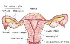
Behind the labia majora and labia minora is the vestibule of the vagina, behind which is the vagina itself. It is a hollow muscular organ located in the pelvis. The vagina occupies a position between the bladder and urethra in front and the rectum in the back. The distal end is attached to the cervix. The cervix is an anatomical formation in the lower segment of the uterus. The cervical canal is an anatomical through hole that passes through the middle of the cervix and directly connects it and the vagina. It may be absent with improper embryogenesis, this condition is called atresia. Normally, the cervical canal is filled with mucus, which protects the uterus from the penetration of microorganisms and other foreign agents.
To understand the essence of a cytological study, it is necessary to understand that organs in different parts of the genital tract are covered with different epithelium. On the surface of the vagina and the vaginal part of the cervix there is a flat stratified epithelium, and in the cervical canal - a cylindrical one. If the cylindrical one goes beyond the canal, this is called ectopia, which is considered a physiological norm and cannot be treated.
Indications for the appointment of a cytological examination of the cervix
The purpose of cervical scraping is to identify abnormal cells and diagnose precancerous diseases. The main indications for the appointment of the study:
Study preparation

The study does not require special preparation, but there are several recommendations that must be followed in order not to get a false result. The study can not be carried out during menstruation. With inflammatory diseases of the genital organs. If at the time of the study a woman experiences pain, itching or burning in the vagina.
IMPORTANT! It is impossible to douche before the study, it is necessary to refrain from sexual intercourse for at least 48 hours and 2 hours before the hemotest, it is necessary to refrain from urinating. If these rules are not observed, the results may be inaccurate or changed.
Carrying out the procedure

First, the obstetrician-gynecologist examines the vagina and cervix in the mirror-dilators and assesses the condition of the mucous membranes; a digital gynecological examination should not be done before the smear. If the epithelium covers a large amount of mucus, it must be removed. Then a scraping is made from the cervix (exocervix), for this, an Eyre spatula is used. After it, a scraping is taken from the cervical canal (), the material is taken using a special cytobrush (Cervix Brash). It is introduced into the canal, 4-5 circular movements are performed. After taking the resulting material is applied to the glass, the smear is dried in air and fixed with alcohol or a special preparation (for Papanicolaou studies). Then the resulting drugs are placed in a container and delivered to the laboratory. If liquid oncocytology is performed, the brush is immersed in a liquid fixative, rinsed, and the brush tip is removed and left in the fixative.
The next stage of the study is laboratory. Laboratory assistants register received samples. Then smears are stained with special dyes (according to Leishman). Liquid cytology preparations are centrifuged or filtered.
Finished preparations are sent to the analytical stage, which is carried out using microscopy. The evaluation criteria are:
- cell type;
- Cell sizes;
- Inclusions in cells;
- Maturity;
- Features and changes of kernels;
- Cytoplasm.
After evaluating the results, the laboratory issues a conclusion, which is sent to the attending physician.
Deciphering the results
Classification of cytological changes according to Papanicolaou:
- Grade 1 - a negative result (normal - there are no atypical cells, the cells have the same shape and size);
- Grade 2 - morphological changes were found that appeared under the influence of inflammation of the vagina or cervix;
- Grade 3 - there is a suspicion of a malignant process, single cells with morphological abnormalities were found;
- class 4 - individual cells with malignant changes;
- Grade 5 - signs of malignant tissue changes were found.
What can a cytological examination show?

- Normal result - no altered cells, phenomena or bacterial vaginosis are possible. You can find unchanged epithelial cells, a moderate number of neutrophils, leukocytes and bacteria;
- Detection of indeterminate atypical cells - such changes cause sexually transmitted infections, HPV, dysplasia, postmenopausal atrophy of the mucosal surface. It is necessary to pass an analysis for the presence of HPV and undergo cytology again in a year;
- A low degree of change in the squamous epithelial cover - dysplasia or HPV infection is possible. The recommendations are the same;
- The presence of atypical cells - the degree or the beginning of the malignant process. For further diagnosis, it is carried out (examination of the walls of the vagina and the visible part of the cervix using a special optical device);
- A high degree of squamous changes - a high level of dysplasia, possibly the uterus. It is necessary to perform a colposcopy, tissue biopsy, if the woman is over 25 years old, it is possible to perform a diagnostic excision (removal of part of the mucosa with further histology of the tissue);
- The presence of atypical cells - epithelial dysplasia of 1-3 degrees, cancer of the cervix or endometrium. Recommendations - colposcopy, diagnostic curettage of the uterus and cervical canal of the uterus, HPV analysis;
- Adenocarcinoma in situ (in situ), squamous cell carcinoma - a high degree of dysplasia or cancerous changes in the cervix. Colposcopy, diagnostic curettage of the uterus and cervical canal of the uterus, HPV analysis are prescribed;
- Benign glandular changes - endometrial hyperplasia. If a woman does not have non-menstrual bleeding before the period, or other pathological processes, then such changes can be attributed to the norm.
Microbiological examination of the cervix and cervical canal
When conducting a cytological study, it is possible to simultaneously conduct microbiological diagnostics. Based on it, a definitive diagnosis cannot be made, but infectious diseases of the genital tract can be suspected.
- Trichomonas colpitis - upon detection of Trichomonas;
- Candidiasis (better known as thrush) - upon detection of fungi of the genus Candida;
- Bacterial vaginosis - a decrease in lactoflora (the normal flora of the vagina), the detection of cocci, gonococci, rods or mixed flora;
- Chlamydia - found chlamydia;
- under the influence of HPV.
To make a final diagnosis, additional studies are needed:
- Microbiological method - flora with subsequent determination of the type of pathogen and its sensitivity to antibacterial drugs;
- PCR - diagnostics (polymerase chain reaction) is a modern diagnostic method based on the determination of the DNA of pathogens of infectious diseases.
Additional methods for diagnosing malignant neoplasms of the female reproductive system

In addition to oncocytology, there are other studies to confirm tumor diseases of the female reproductive system. These include:
- - carried out using an ultrasound machine, allows you to detect changes in the pelvic organs;
- Hysterosalpingography (HSG) is a method of examining the uterus and fallopian tubes, in which their cavities are filled with a contrast agent and X-ray or ultrasound is performed. Allows you to detect obstruction and structural changes in organs;
- Hysteroscopy is an endoscopic examination of the uterine cavity. The advantage of the method is that it can go from diagnostic to therapeutic (allows you to perform minor surgical interventions, for example, or a biopsy);
- Immunohistochemical analysis - a laboratory method for determining the necessary cells, using labeled antibodies;
- Determination of tumor markers in the blood - substances that secrete tumor cells and are not found in the norm.
Conclusion
Oncocytology of the cervix and cervical canal is an important diagnostic method in oncogynecology, although it is not the only one. The assay has many applications, including mass screening of healthy women. The method is simple, has no contraindications and can be performed in outpatient practice. All these advantages have allowed cytological research to take a leading position in medical practice. Thank you for your attention.
Video: cytological and histological examination
Video: epithelial cytology - introduction
Cytological examination (cytology) is the main method of screening assessment of the state of the epithelium of the cervix. The main task of cytological screening is to search for altered epithelial cells (atypical, having a structure different from normal epithelial cells).
The term "abnormal cells" includes both cells with signs of dysplasia - mild, moderate or severe (precancerous cells), as well as cancer cells themselves. The difference between them is in the degree of manifestation of changes in the structure of cells.
Cytological screening must be performed for all women (excluding virgins and patients who have undergone extirpation (removal) of the uterus), starting at 21 years old and ending at 69 years old (in the absence of changes in the studies), the regularity of the analysis is 1 time per year, according to order 572n ( November 1, 2012), however, it is permissible to take an analysis once every three years (MZRF order No. 36 en, dated February 3, 2015).
Currently, there are two alternative methods of fixation and examination of biological material, the key difference between which for patients is their effectiveness.
Pap test and liquid cytology
The sampling of the material is carried out in the same way (standardized sampling): with a combined brush or two cytological brushes (Figure 1), since the epithelium must be taken both from the outer vaginal surface of the cervix (ectocervix) and from the inner one - from the cervical canal (endocervix). The need to take cellular material from the cervical canal is due to the fact that the epithelial junction zone (cylindrical and stratified squamous non-keratinizing - the place where “bad” processes most often begin (90-96% of cases)) moves closer to the center and inside the cervical canal with age.
It is recommended to take cytological material before bimanual (two-handed) vaginal examination, colposcopy and ultrasound. You should not take smears in the presence of vaginitis (an inflammatory process in the vagina), during its treatment, during menstruation. Sexual abstinence is also required for two days.
Biomaterial sampling technique:
- the patient lies on the gynecological chair;
- a mirror is inserted into the vagina, visualizing the cervix;
- the area of \u200b\u200bthe external pharynx is gently blotted with a cotton swab to remove mucus;
- when using two cytobrushes: the first brush is placed on the vaginal surface of the cervix and in the exocervix and rotated 360⁰ clockwise 5 times, and the second brush is placed in the cervical canal at a depth of about 2 cm and rotated at least 3 times counterclockwise;
- when using a combined cytobrush: the central part of the brush, which has short bristles located horizontally, is inserted into the cervical canal, while the long bristles are located on the vaginal part of the cervix, the brush is turned clockwise 3-5 times.
Differences between Pap test and liquid cytology
- In the case of performing a traditional cytological examination (PAP test), the resulting material is distributed on a pre-defatted glass slide in a uniform thin layer, which is not always possible due to the presence of the human factor (the micropreparation is made directly by a specialist), as well as in the presence of an inflammatory process or bloody discharge (epithelial cells can often be obscured by heaps of leukocytes and erythrocytes and are not visible under a microscope). In view of the above, 10% of smears will be non-informative, which will require re-analysis. In addition, most of the collected cells remain on the cytobrushes, for additional studies (in case of obtaining a doubtful result), a second sampling will be necessary.
- The sensitivity of the PAP test (the probability of reliably detecting "unhealthy" cells) is 55-74%, and the specificity (the guarantee that "unhealthy" cells will be detected when they are present in the smear) is 63.2 - 99.4%. The method of liquid cytology has a number of advantages over traditional research.
- When conducting liquid oncocytology, the material is always taken with a combined cytological brush, the collected material, together with a removable brush, is placed in a special container (vial) filled with a stabilizing solution, which prevents the loss of the biomaterial and ensures its long-term storage and additional studies if necessary.
- The information content of liquid cytology is higher, which is ensured by an automatic system for the preparation and staining of micropreparations, which makes it possible to arrange epithelial cells in one layer, separating them from other cellular elements. Preparations are also evaluated automatically using the CytoScreen system.
- The number of inadequate smears when using the liquid technique is 10 times lower than when using the traditional one and does not exceed 1%.
- The biological material remaining as a result of liquid oncocytology can subsequently be used for additional studies, for example, immunocytochemical determination of p16(INK4α) protein or determination of highly oncogenic types of human papillomaviruses.
In 99% of cases, the result obtained using liquid cytology coincides with the results of histological examination.
The only drawback of the method is that it is not included in the compulsory health insurance system, i.e. the analysis is paid.
Cervical cytology results
According to the current clinical guidelines of 2017, the interpretation of the results of the analysis should be carried out according to the Bethesda system, although you can find a cytological conclusion according to the Papanicolaou, WHO and CIN (histological classification) systems. Comparison of systems is shown in table 1.


Conditions defined by the Bethesda terminology system will have clinical significance, therefore, for example, moderate dysplasia, severe dysplasia and carcinoma in situ = CIN II and CIN III = HSIL, and the tactics of managing all of these conditions of stratified squamous epithelium will be the same (HSIL category).
Deciphering the results
So, you are holding an oncocytological conclusion in your hands. The interpretation of the result, as well as the choice of management tactics based on it (taking into account age and lifestyle characteristics), should be carried out not by you, but by your doctor! It is he who directs you to the necessary additional studies and chooses the tactics of treatment, if necessary. But who among us does not look on the Internet to see what the abbreviations made in the cytological conclusion mean and what to prepare for? I think any person worried about their health.
Below we consider the decoding of the abbreviations of the Bethesda terminological system with an indicative (according to the current clinical guidelines (2017) management tactics.
If squamous cells are changed:
NILM
NILM(negative for intraepithelial lesion or malignancy) - negative for dysplasia or cancer- this is the norm, which completely excludes the possibility of the presence of atypical (altered, with signs of possible malignancy) cells. If you see the abbreviation NILM in your conclusion, congratulations! There is a subdivision of a smear negative for dysplasia or cancer into NILM 1 and NILM 2, meaning the first is the absolute norm, and the second is concomitant reactive (inflammatory) changes in the smear, which may be due, for example, to bacterial vaginosis. To clarify the reason for the abbreviation NILM 2 in the cytological conclusion, a smear for flora or PCR diagnostics will help. With regard to the cytological study, there is nothing to worry about. Routine (usual) screening is indicated according to age: at least once every three years up to 29 years; at least once every 5 years in combination with HPV testing over the age of 29; and at least once every 5 years in combination with HPV testing over the age of 29 years.
ASC US
ASC US(atypical squamous cells of undetermined significance, squamous epithelial cells with atypia of unknown significance) is the most common of the variants of deviations found in cytological conclusions. The bottom line is that cells were found that differ in their structure from normal ones, but it is impossible to argue that the differences are due precisely to dysplasia, and not to other causes - reactive states (inflammatory process, hypoestrogenism). According to statistics, the histological picture of CIN III (severe dysplasia) with this cytological conclusion occurs no more often than in 2% of cases. Therefore, you should not worry. But it is worth conducting an HPV test (it should be noted that ASC-US is not accompanied by HPV infection in only one third of cases), and if it is negative, live in peace, having passed both analyzes (oncocytology and HPV testing) in 1-3 years .If HPV is detected, the doctor will prescribe a colposcopy for you, according to the results of which it is possible to take a biopsy (a piece of cervical tissue). In the absence of colposcopic changes in a year, it will be necessary to repeat the cytological examination and HPV testing. Another tactic is also possible: repeated cytological examination in a year. If the conclusion "ASC-US" is received again - colposcopy and HPV - testing, and if "NILM" - no additional studies are required and you can live in peace until the next screening.
ASC-H
ASC-H (atypical squamous cells, cannot exclude HSIL, squamous cells with atypia of unknown significance do not exclude HSIL)- Altered cells were also found here, but the probable cause of their appearance is dysplasia. The doctor will prescribe you a colposcopy with a biopsy and HPV testing, further tactics will be determined depending on the results obtained.
LSIL
HSIL
HSIL(high grade squamous intraepithelial lesion, high-grade squamous intraepithelial lesion) – atypical cells were found in the smear, corresponding to severe dysplastic changes. The doctor will refer you to a colposcopic examination and excision (excision of the area of the altered tissue with a loop) / conization (removal of the cone-shaped portion of the cervix including the vaginal surface and the lower part of the cervical canal) followed by a histological examination of the obtained biomaterial. Category HSIL by Bethesda classifications also includes carcinoma in situ (see Table 1, WHO descriptive system).
CIS
However, cytological examination does not give an idea of the spatial arrangement of cells with signs of atypia; only a histological examination allows to establish the depth of penetration of the pathological process into tissues.
If the cells of the cylindrical epithelium are changed:
AGC
AGC (atypical glandular cells, atypical glandular epithelial cells)- the presence in the collected material of altered cells of the cylindrical epithelium, which in most cases means the location of the pathological process inside the cervical canal.
AIS
AIS (adenocarcinoma in situ, endocervical adenocarcinoma "in situ" (from Latin - "in place")) - the presence of malignantly altered cells of the cylindrical epithelium. As in the case of CIS, it is assumed that the pathological process does not extend beyond the epithelium and the basement membrane is not damaged. But we remember that oncocytological examination cannot determine the depth of tissue damage. It only assesses the level of malignant changes in cells, according to which it determines the degree of dysplasia or determines atypical cells as malignant. In addition to HPV testing and colposcopy, the attending physician will prescribe you a curettage of the cervical canal, and if you are over 35 years old, then an aspiration biopsy of the endometrium to obtain a histological conclusion about the depth of the lesion and exclude the pathological process in the uterine cavity.
The above management tactics, depending on the results of cytological studies, are indicative. The tactics of management in each case is determined by the attending physician, taking into account the individual characteristics of the patient (age, presence or absence of children, concomitant diseases, the fact of HPV infection, personal qualities).
Dear girls, women, I urge you to regularly conduct a cytological examination and wish to receive exclusively "NILM" in conclusion.
Description
Method of determination Microscopy
Material under study See in the description
Home visit available
The vaginal part of the cervix - ectocervix is lined with stratified squamous non-keratinized epithelium. In women of reproductive age, it is constantly rebuilt by proliferation-maturation-desquamation and is completely replaced by a new population of cells every 4 to 5 days.
Normally, the squamous epithelium is represented by the following types of cells: cells of the superficial layer, cells of the intermediate layer, and cells of the basal-parabasal layer. The cellular composition depends on the presence / absence of the menstrual cycle and its phase. The squamous epithelium performs a protective function.
The cervical canal - endocervix - is lined with a cylindrical mucus-producing epithelium. Cyclic changes in the epithelium of the endocervix are poorly expressed. The main function of the cylindrical epithelium is secretory.
The transformation zone is the area of the junction of the stratified squamous and cylindrical epithelium in women of reproductive age, which basically coincides with the area of the external os. Depending on age and hormonal balance in the body, it can also be located on the vaginal part of the cervix. In women of older reproductive and postmenopausal age, the boundary line is actually localized within the external pharynx. According to statistical data, precancer occurs from the zone of transformation.
Material for research. In the direction for the cytological examination of biological material, clinical data, diagnosis, features and place of obtaining the material, data on the menstrual cycle must be indicated.
Swabs are taken prior to bimanual examination and colposcopy. The instruments used must be sterile and dry, as water and disinfectant solutions destroy cellular elements.
During a preventive examination (cytological screening) of women, it is advisable to obtain cell material from the surface of the vaginal part of the cervix (ectocervix) and the walls of the cervical canal (endocervix), in the presence of pathological changes in the cervix aimingly.
Modified Eyre-type spatulas or Cervix-Brash, Papette brushes are used as a tool for taking material from the cervix during a preventive examination of women. For diagnostic purposes, the material is obtained separately with spatulas from the ectocervix, brushes such as Cytobrash from the endocervix.
Material for cytological diagnosis is obtained in various ways: by aspiration and scraping of the contents of the posterior fornix of the vagina, cervix, or by obtaining an imprint smear. The resulting biological material is applied in a thin layer on a glass slide and dried in air. The glass must be marked with not only the last name / code, but also the place where the cell material was taken (cervix, cervical canal). The markings on the slide and in the direction for cytological examination must correspond to each other.
Please note that in children under 16 years of age, gynecological tests are taken only in the presence of parents. Medical offices do not do cervical scrapings and swabs for pregnant women who are 22 weeks or more, as this procedure can cause complications. If necessary, you can contact your doctor to take the material.
Literature
- Petrova AS Cytological diagnosis of tumors and pretumor processes. Medicine, 1985. - p. 296.
- Prilepskaya VN Diseases of the cervix, vagina and vulva. - M.: MEDpress, 1999. - p. 406.
- Shabalova IP Cytological atlas. Moscow, 2001. p. 116.
Training
Preparation conditions are determined by the attending physician. In women of reproductive age, smears should be taken no earlier than on the 5th day of the menstrual cycle and no later than 5 days before the expected start of menstruation. You should not take cell material for research within 24 hours after sexual intercourse, sanitation of the vagina, introduction of medications into the vagina.
Indications for appointment
Cytological smears should be taken from all women over 18 years of age, regardless of clinical data, once a year. In the presence of clinically pronounced pathological changes in the cervix, the cellular material is taken aimingly. The frequency of cytological examination is determined by a gynecologist (at least 2 times a year). (Order No. 430 "On the approval of instructive and methodological guidelines for organizing the work of a women's clinic" dated April 22, 1981 of the USSR Ministry of Health).
The cytological method of research occupies one of the important places in the diagnosis of diseases of the cervix. Due to its high accuracy, it is one of the leading research methods in the diagnosis of background, precancerous and cancerous processes of various localization.
Advantages of the method:
- painlessness and safety of obtaining cellular material;
- the possibility of studying the pathological focus in dynamics;
- the possibility of diagnosing a malignant neoplasm in the initial stage of development;
- small financial costs.
Disadvantages of the method:
- the impossibility of establishing signs of infiltrative growth (cell, not tissue material is examined).
The specificity of this screening method is 69%. The rate of false-negative smears ranges from 5 to 40%. Inadequate sampling from the endocervix is the most important factor in causing false negative results.
The effectiveness of the cytological research method largely depends on the preanalytical stage: how correctly the cellular material is taken and the smears are prepared.
Interpretation of results
The interpretation of test results contains information for the attending physician and is not a diagnosis. The information in this section should not be used for self-diagnosis or self-treatment. An accurate diagnosis is made by the doctor, using both the results of this examination and the necessary information from other sources: history, results of other examinations, etc.
It should be remembered that the cytological method of research, like any other laboratory research method, does not always provide comprehensive information for making a diagnosis. Only a clinician has the right to make a final diagnosis (based on the study of anamnesis, observation of clinical manifestations and data from the histological method of examination).
The result of a cytological examination of the obtained biomaterial (smears-prints) can be presented by a cytologist in the form of: - description of the cellular composition; - descriptions of the cellular composition and conclusions; - descriptions of the cellular composition and conclusions in a hypothetical form; - descriptions of the cellular composition and recommendations.
The form of the answer depends on a number of reasons: the adequacy of the cellular material (few cells, many elements of blood, mucus), an incorrectly completed referral for a cytological examination: the reason for the examination (clinical diagnosis) was not indicated, the presence / absence of menstruation; it is not indicated where the material came from, the marking in the direction does not correspond to that on the glasses, etc.
Result interpretation
Possibilities of cytological diagnosis of certain diseases of the cervix and options for interpreting the results of a cytological study:
Endocervix. Normally, with correctly obtained cellular material from the transformation zone (ZT) - the junction zone of the squamous and columnar epithelium - cells of the squamous and columnar epithelium are present in the smear without changes. Cytological conclusion: cells of squamous and cylindrical epithelium without features were found in the obtained material. The presence of a small amount of metaplastic epithelial cells is an indication that the material was obtained from ST. In the absence of the above description, the swab was not taken from ST and the patient cannot be said to be at no risk of cervical cancer. Such swabs are commonly seen in postmenopausal women and patients who have undergone cervical treatment that has moved the border line into the cervical canal. Depending on the patient's history, this may be a reason for re-sampling the material.
Clinical diagnosis in the direction of the polyp of the cervical canal, and the corresponding cytological picture allow the cytologist to conclude that the cytogram corresponds to the clinical diagnosis of the polyp of the cervical canal. If there is no clinical diagnosis, and the cellular composition is represented by large clusters of cells of the columnar epithelium, the cytologist gives a descriptive answer with the assumption of hyperplasia of the cells of the columnar epithelium or a polyp of the cervical canal.
Ectocervix. In reproductive age, the normal cellular composition of imprints from the vaginal part of the cervix is represented by squamous epithelial cells, predominantly of a superficial or intermediate type. The wording “in the obtained material cells of the squamous epithelium of the surface layers without features are noted” indicates that the obtained biological material consists of cells of the squamous epithelium of the surface and intermediate layers in various combinations in accordance with the phase of the cycle. At the beginning of postmenopause (normal), cells of the squamous epithelium of the intermediate layer are noted in the smear. In some women, during the whole subsequent life, an intermediate type of smear (squamous epithelial cells of the intermediate layer) is observed, sometimes with the presence of cells of the surface layer, which is apparently associated with the function of the adrenal glands, an active sexual life. The presence in the preparation of squamous epithelium cells of the surface layer (estrogenic type of smear) in the first 5 years of menopause should be alarming in relation to neoplasms of the ovaries, uterine fibroids. Postmenopause is characterized by the presence of cells of the basal-parabasal layer (i.e., deep layers).
Erosion (ectopia) of the cervix. The concept of cervical erosion (true erosion) involves a defect in the cervical mucosa caused by various diseases (syphilis, traumatic injuries, the effects of radiation therapy, cervical cancer, etc.). The term cervical ectopia (pseudo-erosion) means the displacement of a high cylindrical epithelium on the vaginal part of the cervix. Provided that there is a clinical diagnosis of "erosion / ectopia of the cervix" in the direction and the correct sampling of biomaterial from the ectocervix (cellular material is represented by squamous epithelial cells of all layers in various combinations, clusters of cylindrical epithelium cells, elements of inflammation), the cytological conclusion has the following form of answer: the cytogram corresponds (does not contradict) the clinical diagnosis - erosion of the cervix.
Cytological conclusion: the cytogram corresponds (does not contradict) the clinical diagnosis of cervical ectopia suggests the presence in the obtained material of squamous epithelial cells of the surface layers, clusters of cylindrical epithelium cells.
Conclusion: a cytogram of endocervicosis occurs if the clinical diagnosis of erosion/ectopia of the cervix is not indicated in the referral for cytological examination, and morphologically, cells of the squamous epithelium and clusters of cells of the cylindrical epithelium are noted.
It is not always possible to make a cytological diagnosis between superficial endocervicosis (ectopia of the cervix) and proliferating endocervicosis. A descriptive cytological response occurs when: - cells of the squamous epithelium and single clusters or single cells of the cylindrical epithelium are found in the material obtained from the ectocervix; - cellular material obtained from ecto- and endocervix and presented in one mixed smear; - smears are not smeared.
With healing endocervicosis, a large number of cells of metaplastic epithelium are found in smears (metaplasia is the replacement of one type of epithelium with another). Metaplastic epithelium is a target for human papillomavirus exposure - an area for the development of precancerous conditions. The presence in smears from the cervix of a small number of cells of metaplastic epithelium is an indicator of a normal physiological process.
Histogenetic mechanisms of replacement of the columnar epithelium by squamous: - progression of squamous cell transformation - direct ingrowth of the native epithelium under the columnar. As the squamous cells develop and mature, the endocervical cells move upward, degenerate, and eventually slough off. A similar process is observed during re-epithelialization of true cervical erosion healing; - squamous metaplasia - proliferation of undifferentiated reserve cells of the endocervical epithelium and their partial transformation into a fully mature squamous epithelium. The first stage of the process is the appearance of reserve cells, then comes reserve cell hyperplasia, followed by differentiation into immature squamous epithelium, and at the final stage mature squamous epithelium is observed.
Leukoplakia of the cervix. With the cytological method for diagnosing simple leukoplakia (a benign lesion of the cervix, a background disease), hyperkeratosis is detected, i.e., in the material obtained from ectocervix, layers (clusters) of squamous epithelium scales were found (there is no nucleus in the cytoplasm of the cell), separately lying squamous epithelium scales, dyskerocytes . If there is a clinical diagnosis of "leukoplakia of the cervix" - in the cytological report it is noted that the picture does not contradict the clinical diagnosis - leukoplakia of the cervix. In the absence of a clinical diagnosis of cervical leukoplakia, depending on the available material, the cytologist gives a descriptive answer, possibly with a recommendation to exclude cervical leukoplakia. Single scales of squamous epithelium have no diagnostic value. Leukoplakia with atypia - a cytological method of research is not always possible to identify, which is explained by the presence of squamous epithelium scales on the surface of the stratified squamous epithelium, which prevent the receipt of cellular elements. It is necessary to conduct a morphological study of the biopsy of the cervix.
Dysplasia of the cervix. Dysplastic changes occur in the stratified squamous epithelium of both exocervix and endocervix. As a rule, changes begin at the junction of the squamous and columnar epithelium. Dysplasia can simultaneously develop in several parts of the cervix and cervical canal, often changes are expressed to varying degrees. Spectrum dysplasia (CIN) is not a single disease. There are two biological entities of the process: a productive human papillomavirus infection and a cancer precursor.
Dysplasia-I (mild dysplasia, CINI) is one of the least reproducible cytological diagnoses. Dysplasia-I is often difficult to differentiate from reactive epithelium. It is not always possible to make a differential diagnosis between dysplasia III (severe dysplasia, CIN-III) and intraepithelial cancer by cytological examination.
Cytological conclusion: Dysplasia - I (weak, CIN-1); Dysplasia -II (moderate, CIN-II); Dysplasia -III (severe, pronounced, CIN-III). If there are cells with signs of malignancy in the obtained material, the cytologist gives a conclusion on the cytogram of the malignant neoplasm and, if possible, specifies the form of cancer.
Inflammatory processes of the cervix. Inflammation - a cellular reaction (in the focus) - is represented by a degeneratively altered epithelium, proliferative changes of a reparative, protective nature, and inflammatory atypia. In an acute nonspecific inflammatory process, a pronounced leukocyte infiltration (many neutrophilic leukocytes), incomplete phagocytosis is noted in the smear. The composition of the cell population of the epithelium may change. Cytological conclusion: cytogram of ecto-/endocervicitis. In subacute and chronic inflammation, eosinophils, lymphocytes, macrophages/cells such as foreign bodies (multinuclear macrophages) join - cytological conclusion: a cytogram of chronic ecto-/endocervicitis. Acute inflammatory processes are more often observed in the age group of 20-24 years, chronic processes and their consequences occur in women aged 25-34.
Infectious lesions of the cervix. Cytological features of smears for infectious lesions of the cervix depend on the pathogen and the duration of the inflammatory process.
Mycoplasmas, ureaplasmas and corynobacteria as the cause of inflammation are observed in the group of young women (up to 20 years old). In the age group over 30 years, anaerobic microorganisms occupy the first place among the causative agents of inflammatory processes in the genitals. Mixed infection increases the pathogenicity of each of the pathogens. In such cases, inflammation causes a pronounced tissue reaction, accompanied by damage to the epithelium, destruction and dysplasia. This leads to the development of not only colpitis, endocervicitis, but can play a significant role in the formation of cervical ectopia. Incomplete phagocytosis is noted (phagocytic activity of leukocytes is suppressed). The cytological conclusion indicates the type of flora with a recommendation to exclude a certain type of infection.
Bacterial vaginosis (BV) - (clinical diagnosis). In cytological preparations, BV is represented by key cells. If the key cells are not found, and the flora is cocco-bacillary, it is recommended to exclude the presence of gardnerella (ureaplasma) in the cytological response; in the presence of mobiluncus bacilli, a recurrence of the pathological process after the treatment is possible.
Genital herpes - the herpes simplex virus has a high tropism for epithelial and nerve cells. Relapses are mainly due to the persistence of infection in the nerve ganglion. Cytological examination of the obtained material may show changes in squamous epithelial cells specific for their defeat by this type of viral infection: multinucleated cells of the "mulberry" type. Form of cytological response: signs of a viral infection were found in the material obtained. It is recommended to exclude the herpes simplex virus.
Papillomavirus infection of the genitals. The human papillomavirus is able to persist for a long time in the basal layer of the squamous epithelium, which causes a high frequency of recurrence of the process. The frequency of coincidence of cytological and histological diagnoses in condyloma was 42%: CIN-I - 56%, CIN III 74%. False-negative cytological responses are explained by the consequence of incorrect material sampling - 90%, incorrect interpretation - 10%.
In addition, underdiagnosis in cervical smears may be due to the presence of koilocytes in the deeper layers of the squamous epithelium or the presence of a large overlap of elements of inflammation and flora. Cytological conclusion: the obtained material showed signs of a viral infection. It is recommended to rule out the human papillomavirus. Indirect changes characteristic of a viral infection: an increase in the size of the nucleus, nonspecific multinucleation. The form of the cytological response: the obtained material shows indirect signs of a viral infection. It is recommended to exclude the herpes simplex virus, human papillomavirus.
Trichomoniasis. An inflammatory reaction develops in the presence of a large number of protozoa. Proper preparation of the patient is essential for the quality of the study. Termination of the use of trichomonocidal drugs for 5-7 days before taking the material. In the cytological preparation, there are signs of an acute/chronic inflammatory process, mixed flora, Trichomonas. Cytological conclusion: trichomonas colpitis.
Chlamydial infection. Chlamydia are tropic to columnar epithelium. Often found in women with cervical ectopia. In pregnant women and menopausal women, signs of infection may be observed in the squamous epithelium. They can also be found in macrophages. Cytologically, the presence of intracellular specific inclusions is determined, which are more often detected with a fresh or untreated infection. Cytological forms of response: cells with cytoplasmic inclusions morphologically similar to chlamydial infection were found. It is recommended to exclude the presence of chlamydial infection.
Squamous intraepithelial lesions (SIP) of the cervix are associated with significant qualitative and quantitative changes in the vaginal microflora. Deficiency of lactobacilli is observed in all patients with PIP, there is an increase in representatives of opportunistic flora. In the cytological conclusion, changes in the flora are indicated, if possible, a representative of the opportunistic flora is characterized. The presence of nonspecific vaginosis is noted.
Women's health requires the constant intervention of narrow medical specialists. When a woman is pregnant, she seeks advice from a gynecologist; when she gives birth, midwives come to the rescue. During medical examinations, the fair sex must be examined by mammologists and the same gynecologists. Health is priceless, that's why we care about it so much. Recently, such a disease as cancer destroys more and more bright hopes for a wonderful future. Oncology of the uterus or mammary glands is dangerous because in the first stages it cannot be determined if you do not periodically come for examinations.
The Science of Cytology to Help Diagnosis
Cytology is not fully related to the medical sciences. Rather, it is more biological, but is important for the diagnosis of various diseases. This science is concerned with the study of the structure and basic functions of living cells. Under the microscope, the entire cycle of cell existence is determined. From its inception to aging and death. Particular importance is given to the reproduction of living cells, the presence of organelles, the occurrence of any pathological processes in their functioning.
Medicine actively uses the developments of this science for its diagnostic purposes. To date, cytological studies of scrapings from the cervix are widely used. Knowledge of the structure and structure of cells makes it possible to develop innovative technologies in the treatment of dangerous diseases. Cytology has become a branch of laboratory research. It does not make any predictions, but is only descriptive. Oncocytology has become a new section - a science that helps diagnose neoplasms as soon as they appear.
Cytological examination in gynecology
With pathologies of the cervix or suspicion of them, a cytological examination of the smear is performed. Before the start and end of the treatment of gynecological diseases, as well as during the usual planned medical examination, a smear for cytology is mandatory. This study assesses the condition of the cells of the cervix and other female organs.

For the first time such an analysis was carried out in the thirties of the last century. And the first classification of cells taken for cytological examination was published in 1954. It was changed several times, and its present version was developed in 1988. According to this version, cervical cells are divided into different classes, characterizing the degree of atypicality, ranging from normal to invasive cancer. These data are of great diagnostic value and allow you to choose the most effective therapy.
Examination of cervical cells with a smear
A smear is not taken during a colposcopy or vaginal examination. The procedure itself is carried out under a microscope. Epithelial cells tend to be constantly updated, that is, to exfoliate. They appear in the lumen of the cervix and in the vagina. The structure of these cells is such that both healthy and atypical elements can be determined by microscopy.

One of the simplest and least invasive research methods, which is not accompanied by discomfort, is the pap test. This procedure allows you to detect the possibility of degeneration of cervical cells into cancer.
Also, with the help of this test, it is possible to diagnose a tumor process in other female organs, for example, in the uterus or ovaries. Unfortunately, the Pap test is not always accurate. There were situations when, after several negative results, a woman was still diagnosed with cervical cancer. But, perhaps, such incidents happened due to the wrong taking of the material. Malignant degeneration begins from the lower layers and gradually grows upward. If you take only the surface layer, then you can only notice malignant changes at the final stage.
Scraping for cytological examination
The material for a cytological examination of a smear is taken with a brush and a special spatula, with which cells arranged in layers are scraped off with pressure. During this procedure, a lot of material from the cervix gets on the glass, the structure of which does not change.

This process is completely painless. The cells are scraped off in several places and put on a glass slide. After that, the preparation is fixed with a special solution and stained with dyes. The smear is then sent for testing.
The result of a cytological study may indicate the presence of atypical cells that occur with severe inflammation or cancer.
How is the study of cervical cells carried out?
Cryocautery is an absolutely safe and painless procedure. It consists in the fact that the affected areas of the tissues of the cervix are frozen with a special probe. Then they peel off.
Also, after a cytological examination of the cervix using scraping, the gynecologist may prescribe additional procedures such as laser therapy and excision of the pathological area with a loop.