Molecular biology and biological chemistry study. Subject, tasks and goals of molecular biology. b). additional literature
interview
Pirogov Sergey - a participant in the preparation for the Olympiad in biology, organized by "Elephant and Giraffe" in 2012.
Winner of the International Universiade in Biology
The winner of the Olympiad "Lomonosov"
Regional stage winner All-Russian Olympiad in biology in 2012
Studying at Moscow State University. M.V. Lomonosov at the Faculty of Biology: Department of Molecular Biology, 6th year student. Works in the Laboratory of Biochemical Genetics of Animals of the Institute of Molecular Genetics.
- Seryozha, if readers have questions, will they be able to ask you?
Yes, of course, you can ask questions at least immediately. In this field:
Click here to ask a question.

- Let's start with school, didn't you have a super-cool school?
I studied at a very weak Moscow school, such an average secondary school. True, we had a wonderful teacher at the Moscow Art Theater, thanks to whom we had a largely nominal "art history" orientation of the school.
- What about biology?
Our biology teacher was a very elderly, deaf and sharp woman, whom everyone was afraid of. But love for her subject did not add. I have been passionate about biology since childhood, from the age of five. I read everything myself, mainly being carried away by anatomy and zoology. So school subjects existed in parallel with my own interests. The Olympics changed everything.
- Tell me more about it.
In the 7th grade, I took part in the municipal stage for the first time (of course, in almost all subjects at once, since I was the only student whom the teachers had reason to send). And he won in biology. Then the school treated this as a funny, but not very interesting fact.

- Did it help you in school?
I remember that despite my brilliant studies, I often received B from a biology teacher with nit-picking like "in the drawing of a section of an onion, the roots should be painted brown, not gray." It was all pretty depressing. In the 8th grade, I again went to the Olympiad, but for some reason I was not sent in biology. But he became a winner and prize-winner in other subjects.
- What happened in 9th grade?
In the 9th grade, I did not go to the district stage. It was there that I unexpectedly scored a weak, borderline score, which nevertheless turned out to be passing to the regional stage. It had a powerful motivating force - the realization of how much I don’t know and how many people who know all this (how many such people on a national scale I was even afraid to imagine).

- Tell us how you prepared.
Intensive self-study, forays into bookstores, and thousands of last year's assignments had a healing effect. I scored one of the highest scores for the theory (which was also completely unexpected for me), went to the practical stage ... and failed it. At that time, I did not even know about the existence of the practical stage.
- Did the Olympics influence you?
My life has changed radically. I learned about many other Olympiads, especially I fell in love with the SBO. Subsequently, he showed good results on many, won some, thanks to Lomonosovskaya he received the right to enter without exams. At the same time, I won Olympiads in the history of art, to which I still breathe unevenly. True, he was not friends with practical tours. In the 11th grade, I nevertheless reached the final stage, but Fortune was not favorable, and this time I did not have time to fill out the answer matrix of the theoretical stage. But this made it possible not to worry too much about the practical.
- Have you met many Olympiads?
Yes, I still think that I was very lucky with the circle of my peers, who greatly expanded my horizons. The other side of the Olympiads, in addition to the motivation to study the subject more harmoniously, was acquaintance with the Olympiads. Already at that time, I noticed that horizontal communication is sometimes more useful than vertical communication - with teachers at the training camp.

- How did you enter the university? Did you choose a faculty?
After the 11th grade, I entered the Faculty of Biology of Moscow State University. Just the majority of my then comrades made a choice in favor of the FBB, but here the primary role was played by the fact that I did not become the winner of the All-Russian. So I would have to take an internal exam in mathematics, and in it, especially at school - I fell in love with the higher one much more - I was not strong. And there was very poor preparation at school (we were not even prepared for almost the entire C part). In terms of interests, even then I guessed that, in the end, you can come to any result, regardless of the place of admission. Subsequently, it turned out that there are many FBB graduates who switched to predominantly wet biology, and vice versa - many good bioinformaticians started out as amateurs. Although at that moment it seemed to me that the contingent at the biological faculty would be unlike the FBBshny one. In this I was certainly wrong.
Did you know?
interesting
Did you know?
interesting
In the camp Elephant and Giraffe there are shifts in biochemistry and molecular biology, where schoolchildren, together with experienced teachers from Moscow State University, set up experiments and also prepare for Olympiads.© Interviewed by Reshetov Denis. The photos were kindly provided by Sergey Pirogov.
It can be said that molecular biology explores the manifestations of life on inanimate structures or systems with elementary signs of vital activity (which can be individual biological macromolecules, their complexes or organelles), studying how the key processes that characterize living matter are realized through chemical interactions and transformations.
The separation of molecular biology from biochemistry into an independent field of science is dictated by the fact that its main task is to study the structure and properties of biological macromolecules involved in various processes, to elucidate the mechanisms of their interaction. Biochemistry, on the other hand, deals with the study of the actual processes of vital activity, the patterns of their course in a living organism, and the transformations of molecules that accompany these processes. Ultimately, molecular biology tries to answer the question of why this or that process occurs, while biochemistry answers the questions of where and how, from the point of view of chemistry, the process in question occurs.
History
Molecular biology as a separate area of biochemistry began to take shape in the 1930s. It was then that for a deeper understanding of the phenomenon of life, the need arose for targeted studies at the molecular level of the processes of storage and transmission of hereditary information in living organisms. Then the task of molecular biology was defined in the study of the structure, properties and interaction of nucleic acids and proteins. The term "molecular biology" was first used by the English scientist William Astbury in the context of research related to elucidating the relationship between the molecular structure and the physical and biological properties of fibrillar proteins, such as collagen, blood fibrin, or muscle contractile proteins.
In the early days of molecular biology, RNA was considered a component of plants and fungi, while DNA was seen as a typical component of animal cells. The first researcher to prove that DNA is found in plants was Andrey Nikolaevich Belozersky, who isolated pea DNA in 1935. This discovery established the fact that DNA is a universal nucleic acid present in plant and animal cells.
A major achievement was the establishment by George Beadle and Edward Tatum of a direct causal relationship between genes and proteins. In their experiments, they exposed neurospore cells ( Neurosporacrassa) X-ray exposure that caused mutations. The results obtained showed that this led to a change in the properties of specific enzymes.
In 1940, Albert Claude isolated cytoplasmic RNA-containing granules from the cytoplasm of animal cells, which were smaller than mitochondria. He called them microsomes. Subsequently, in the study of the structure and properties of the isolated particles, their fundamental role in the process of protein biosynthesis was established. In 1958, at the first symposium dedicated to these particles, it was decided to call these particles ribosomes.
Another important step in the development of molecular biology was the published data of the experiment of Oswald Avery, Colin MacLeod and MacLean McCarthy in 1944, which showed that DNA is the cause of bacterial transformation. This was the first experimental evidence of the role of DNA in the transmission of hereditary information, debunking the earlier idea of the protein nature of genes.
In the early 1950s, Frederick Sanger showed that a protein chain is a unique sequence of amino acid residues. In the late 1950s, Max Perutz and John Kendrew deciphered the spatial structure of the first proteins. Already in 2000, hundreds of thousands of natural amino acid sequences and thousands of spatial structures of proteins were known.
Around the same time, Erwin Chargaff's research allowed him to formulate rules describing the ratio of nitrogenous bases in DNA (the rules say that regardless of species differences in DNA, the amount of guanine is equal to the amount of cytosine, and the amount of adenine is equal to the amount of themin), which later helped to make the greatest a breakthrough in molecular biology and one of the greatest discoveries in biology in general.
This event occurred in 1953 when James Watson and Francis Crick, based on the work of Rosalind Franklin and Maurice Wilkins on X-ray diffraction analysis DNA, established the double-stranded structure of the DNA molecule. This discovery made it possible to answer the fundamental question about the ability of the carrier of hereditary information to self-reproduce and to understand the mechanism of transmission of such information. The same scientists formulated the principle of complementarity of nitrogenous bases, which is of key importance for understanding the mechanism of formation of supramolecular structures. This principle, which is now used to describe all molecular complexes, makes it possible to describe and predict the conditions for the emergence of weak (nonvalent) intermolecular interactions, which determine the possibility of the formation of secondary, tertiary, etc. structures of macromolecules, self-assembly of supramolecular biological systems that determine such a wide variety of molecular structures and their functional sets. Then, in 1953, arose Science Magazine Journal of Molecular Biology. It was headed by John Kendrew, whose area of scientific interest was the study of the structure of globular proteins (Nobel Prize in 1962, jointly with Max Perutz). A similar Russian-language journal called Molecular Biology was founded in the USSR by V. A. Engelhardt in 1966.
In 1958, Francis Crick formulated the so-called. the central dogma of molecular biology: the idea of the irreversibility of the flow of genetic information from DNA through RNA to proteins according to the scheme DNA → DNA (replication, creation of a copy of DNA), DNA → RNA (transcription, copying of genes), RNA → protein (translation, decoding of information about the structure proteins). This dogma was somewhat corrected in 1970, taking into account the accumulated knowledge, since the phenomenon of reverse transcription was discovered independently by Howard Temin and David Baltimore: an enzyme was discovered - reverse transcriptase, which is responsible for the implementation of reverse transcription - the formation of double-stranded DNA on a single-stranded RNA template, which occurs in oncogenic viruses. It should be noted that the strict necessity of the flow of genetic information from nucleic acids to proteins still remains the basis of molecular biology.
In 1957, Alexander Sergeevich Spirin, together with Andrei Nikolaevich Belozersky, showed that, despite significant differences in the nucleotide composition of DNA from different organisms, the composition of total RNA is similar. Based on these data, they came to the sensational conclusion that the total RNA of a cell cannot act as a carrier of genetic information from DNA to proteins, since it does not correspond to it in its composition. At the same time, they noticed that there is a minor fraction of RNA, which fully corresponds in its nucleotide composition to DNA and which can be a true carrier of genetic information from DNA to proteins. As a result, they predicted the existence of relatively small RNA molecules, which are analogous in structure to individual sections of DNA and act as intermediaries in the transfer of genetic information contained in DNA to the ribosome, where protein molecules are synthesized using this information. In 1961 (S. Brenner, F. Jacob, M. Meselson on the one hand and F. Gros, Francois Jacob and Jacques Monod were the first to experimentally confirm the existence of such molecules - informational (matrix) RNA. At the same time they developed the concept and model of functional units of DNA - an operon, which made it possible to explain exactly how the regulation of gene expression in prokaryotes is carried out. The study of the mechanisms of protein biosynthesis and the principles of the structural organization and operation of molecular machines - ribosomes - made it possible to formulate a postulate describing the movement of genetic information, called the central dogma of molecular biology: DNA - mRNA is a protein.
In 1961 and over the next few years, Heinrich Mattei and Marshall Nirenberg, and then Har Korana and Robert Holly, carried out several works to decipher the genetic code, as a result of which a direct relationship was established between the DNA structure and synthesized proteins and the nucleotide sequence that determines set of amino acids in a protein. Data on the universality of the genetic code were also obtained. The discoveries were awarded the Nobel Prize in 1968.
For the development of modern ideas about the functions of RNA, the discovery of non-coding RNA, made on the basis of the results of the work of Alexander Sergeevich Spirin together with Andrei Nikolayevich Belozersky in 1958, Charles Brenner with co-authors and Saul Spiegelman in 1961, was decisive. This type of RNA makes up the bulk of cellular RNA. Ribosomal RNAs are primarily non-coding.
Methods for cultivating and hybridizing animal cells have received serious development. In 1963, François Jacob and Sydney Brenner formulated the replicon, a sequence of inherently replicating genes that explains important aspects of the regulation of gene replication.
In 1967, in the laboratory of A. S. Spirin, it was demonstrated for the first time that the shape of compactly folded RNA determines the morphology of the ribosomal particle.
In 1968, a significant fundamental discovery was made. Okazaki, having discovered DNA fragments of the lagging strand in the study of the replication process, named Okazaki fragments after her, clarified the mechanism of DNA replication.
In 1970, a significant discovery was made independently by Howard Temin and David Baltimore: an enzyme was discovered - reverse transcriptase, which is responsible for the implementation of reverse transcription - the formation of double-stranded DNA on a single-stranded RNA template, which occurs in oncogenic viruses containing RNA.
Another important achievement of molecular biology was the explanation of the mechanism of mutations at the molecular level. As a result of a series of studies, the main types of mutations were established: duplications, inversions, deletions, translocations and transpositions. This made it possible to consider evolutionary changes from the point of view of gene processes, and made it possible to develop the theory of molecular clocks, which is used in phylogeny.
By the beginning of the 1970s, the basic principles of the functioning of nucleic acids and proteins in a living organism had been formulated. It was found that proteins and nucleic acids in the body are synthesized according to a matrix mechanism, the matrix molecule carries encrypted information about the sequence of amino acids (in a protein) or nucleotides (in a nucleic acid). During replication (doubling of DNA) or transcription (synthesis of mRNA), DNA serves as such a template, during translation (protein synthesis) or reverse transcription - mRNA.
Thus, theoretical prerequisites were created for the development of applied areas of molecular biology, in particular, genetic engineering. In 1972 Paul Berg, Herbert Bauer and Stanley Cohen developed molecular cloning technology. Then they were the first to obtain recombinant DNA in vitro. These outstanding experiments laid the foundations of genetic engineering, and this year is considered the birth date of this scientific direction.
In 1977, Frederick Sanger and independently Allan Maxum and Walter Gilbert developed various methods for determining the primary structure (sequencing) of DNA. The Sanger method, the so-called chain termination method, is the basis of the modern sequencing method. The principle of sequencing is based on the use of labeled bases that act as terminators in a cyclic sequencing reaction. This method has become widespread due to the ability to quickly conduct analysis.
1976 - Frederick. Sanger deciphered the nucleotide sequence of the DNA of the phage φΧ174 with a length of 5375 nucleotide pairs.
1981 - Sickle cell anemia becomes the first genetic disease to be diagnosed by DNA analysis.
1982-1983 the discovery of the catalytic function of RNA in the American laboratories of T. Check and S. Altman changed the existing ideas about the exclusive role of proteins. By analogy with catalytic proteins - enzymes, catalytic RNAs were called ribozymes.
1987 Keri Mullez discovered the polymerase chain reaction, thanks to which it is possible to artificially significantly increase the number of DNA molecules in solution for further work. Today it is one of the most important methods of molecular biology used in the study of hereditary and viral diseases, in the study of genes and in genetic identification and kinship, etc.
In 1990, at the same time, three groups of scientists published a method that made it possible to quickly obtain synthetic functionally active RNAs in the laboratory (artificial ribozymes or molecules that interact with various ligands - aptamers). This method is called "evolution in vitro". And soon after that, in 1991-1993 in the laboratory of A.B. Chetverina was experimentally shown the possibility of existence, growth and amplification of RNA molecules in the form of colonies on solid media.
In 1998, almost simultaneously, Craig Mello and Andrew Fire described the mechanism observed earlier in gene experiments with bacteria and flowers. RNA interference, in which a small double-stranded RNA molecule leads to a specific suppression of gene expression.
The discovery of the mechanism of RNA interference is of great practical importance for modern molecular biology. This phenomenon is widely used in scientific experiments as a tool for "turning off", that is, suppressing the expression of individual genes. Of particular interest is the fact that this method allows reversible (temporary) suppression of the activity of the studied genes. Research is underway to apply this phenomenon to the treatment of viral, neoplastic, degenerative and metabolic diseases. It should be noted that in 2002, mutants of polio viruses were discovered that can avoid RNA interference, so more painstaking work is required to develop a truly effective methods treatment based on this phenomenon.
In 1999-2001, several groups of researchers determined the structure of the bacterial ribosome with a resolution of 5.5 to 2.4 angstroms.
Subject
The achievements of molecular biology in the knowledge of living nature can hardly be overestimated. Great success has been achieved thanks to a successful concept of research: complex biological processes are considered from the standpoint of individual molecular systems, which makes it possible to apply precise physicochemical research methods. It also attracted many great minds from related areas to this area of science: chemistry, physics, cytology, virology, which also had a beneficial effect on the scale and speed of development of scientific knowledge in this area. Such significant discoveries as the determination of the structure of DNA, the deciphering of the genetic code, the artificial directed modification of the genome, made it possible to understand much more deeply the specifics of the developmental processes of organisms and successfully solve numerous important fundamental and applied scientific, medical and social problems that were considered unsolvable not so long ago.
The subject of study in molecular biology is mainly proteins, nucleic acids and molecular complexes (molecular machines) based on them and the processes in which they participate.
Nucleic acids are linear polymers consisting of nucleotide units (compounds of a five-membered sugar with a phosphate group at the fifth atom of the cycle and one of the four nitrogenous bases) interconnected by an ester bond of phosphate groups. Thus, nucleic acid is a pentose phosphate polymer with nitrogenous bases as side substituents. The chemical composition of the RNA chain differs from DNA in that the former consists of a five-membered ribose carbohydrate cycle, while the latter consists of a dehydroxylated ribose derivative, deoxyribose. At the same time, these molecules differ dramatically in space, since RNA is a flexible single-stranded molecule, while DNA is a double-stranded molecule.
Proteins are linear polymers, which are chains of alpha-amino acids interconnected by a peptide bond, hence their second name - polypeptides. The composition of natural proteins includes many different amino acid units - up to 20 in humans, which determines a wide variety of functional properties of these molecules. These or other proteins are involved in almost every process in the body and perform many tasks: they play the role of cellular building material, provide transport of substances and ions, catalyze chemical reactions - this list is very long. Proteins form stable molecular conformations of various levels of organization (secondary and tertiary structures) and molecular complexes, which further expands their functionality. These molecules can have a high specificity for performing certain tasks due to the formation of a complex spatial globular structure. A wide variety of proteins ensures the constant interest of scientists in this kind of molecules.
Modern ideas about the subject of molecular biology are based on a generalization first put forward in 1958 by Francis Crick as the central dogma of molecular biology. Its essence was the assertion that genetic information in living organisms goes through strictly defined stages of implementation: copying from DNA to DNA at the entrance of inheritance, from DNA to RNA, and then from RNA to protein, and the reverse transition is not feasible. This statement was true only in part, therefore, subsequently, the central dogma was corrected with an eye to the newly discovered data.
On the this moment there are several ways to implement the genetic material, representing different sequences of implementation three types the existence of genetic information: DNA, RNA and protein. In nine possible ways of realization, three groups are distinguished: these are three general transformations (general), which are carried out normally in most living organisms; three special transformations (special), carried out in some viruses or in special laboratory conditions; three unknown transformations (unknown), the implementation of which is considered impossible.
Common transformations include the following ways of implementing the genetic code: DNA→DNA (replication), DNA→RNA (transcription), RNA→protein (translation).
To carry out the transfer of hereditary traits, parents need to pass on a full-fledged DNA molecule to their descendants. The process by which an exact copy of the original DNA can be synthesized, and therefore genetic material can be transferred, is called replication. It is carried out by special proteins that unravel the molecule (straighten its section), unwind the double helix and, using DNA polymerase, create an exact copy of the original DNA molecule.
To ensure the life of a cell, it needs to constantly refer to the genetic code embedded in the DNA double helix. However, this molecule is too large and clumsy to be used as a direct source of genetic material for continuous protein synthesis. Therefore, in the course of implementing the information embedded in DNA, there is an intermediary stage: the synthesis of mRNA, which is a small single-stranded molecule complementary to a certain segment of DNA encoding a certain protein. The transcription process is provided by RNA polymerase and transcription factors. The resulting molecule can then be easily delivered to the part of the cell responsible for protein synthesis - the ribosome.
After RNA enters the ribosome, the final stage of the realization of genetic information begins. At the same time, the ribosome reads the genetic code from mRNA in triplets called codons and synthesizes the corresponding protein based on the information received.
In the course of special transformations, the genetic code is realized according to the scheme RNA → RNA (replication), RNA → DNA (reverse transcription), DNA → protein (direct translation). Replication of this type is realized in many viruses, where it is carried out by the enzyme RNA-dependent RNA polymerase. Similar enzymes are also found in eukaryotic cells, where they are associated with the process of RNA silencing. Reverse transcription has been found in retroviruses, where it is carried out by the enzyme reverse transcriptase, and in some cases in eukaryotic cells, for example, during telomeric synthesis. Live transmission is carried out only in artificial conditions in an isolated system outside the cell.
Any of the three possible transitions of genetic information from protein to protein, RNA or DNA is considered impossible. The case of the action of prions on proteins, as a result of which a similar prion is formed, could conditionally be attributed to the type of realization of genetic information protein → protein. However, formally it is not such, since it does not affect the amino acid sequence in the protein.
The history of the emergence of the term "central dogma" is curious. Since the word dogma generally means a statement that is not subject to doubt, and the word itself has a clear religious connotation, choosing it as a description of a scientific fact is not entirely legitimate. According to Francis Crick himself, it was his mistake. He wanted to give the theory put forward more significance, to distinguish it from the background of other theories and hypotheses; why he decided to use this majestic, in his opinion, word, not understanding its true meaning. The name, however, stuck.
Molecular biology today
The rapid development of molecular biology, the constant interest in achievements in this field on the part of society and the objective importance of research have led to the emergence of a large number of large research centers of molecular biology around the world. Among the largest, the following should be mentioned: the laboratory of molecular biology in Cambridge, the Royal Institute in London - in the UK; institutes of molecular biology in Paris, Marseille and Strasbourg, Pasteur Institute - in France; departments of molecular biology at Harvard University and the Massachusetts Institute of Technology, the University of Berkeley, the California Institute of Technology, the Rockefeller University, the Institute of Public Health in Bethesda - in the USA; the Max Planck institutes, the universities in Göttingen and Munich, the Central Institute for Molecular Biology in Berlin, the institutes in Jena and Halle - in Germany; Karolinska Institute in Stockholm, Sweden.
In Russia, the leading centers in this field are the Institute of Molecular Biology. Institute of Molecular Genetics RAS, Institute of Gene Biology RAS, Institute of Physicochemical Biology named after V.A. A. N. Belozersky Moscow State University. M.V. Lomonosov Institute of Biochemistry. A.N. Bach RAS and the Institute of Protein RAS in Pushchino.
Today, the field of interest of molecular biologists covers a wide range of fundamental scientific issues. As before, the leading role is occupied by the study of the structure of nucleic acids and protein biosynthesis, the study of the structure and functions of various intracellular structures and cell surfaces. Also important areas of research are the study of the mechanisms of reception and signal transmission, the molecular mechanisms of the transport of compounds within the cell and also from the cell to the external environment and back. Among the main directions of scientific research in the field of applied molecular biology, one of the most priority is the problem of the emergence and development of tumors. Also a very important area, which is studied by the section of molecular biology - molecular genetics, is the study of the molecular basis of the occurrence of hereditary diseases, and viral diseases, such as AIDS, as well as the development of methods for their prevention and, possibly, treatment at the gene level. The discoveries and developments of molecular biologists in forensic medicine have found wide application. A real revolution in the field of personal identification was made in the 80s by scientists from Russia, the USA and Great Britain thanks to the development and implementation of the method of "genomic fingerprinting" - the identification of DNA in everyday practice. Research in this area does not stop to this day, modern methods make it possible to establish a person with a probability of error of one billionth of a percent. Already, there is an active development of a project of a genetic passport, which, as expected, will greatly reduce the level of crime.
Methodology
Today, molecular biology has an extensive arsenal of methods to solve the most advanced and most complex problems facing scientists.
One of the most common methods in molecular biology is gel electrophoresis, which solves the problem of separating a mixture of macromolecules by size or charge. Almost always, after the separation of macromolecules in the gel, blotting is used, a method that allows you to transfer macromolecules from the gel (sorb) to the membrane surface for the convenience of further work with them, in particular hybridization. Hybridization - the formation of hybrid DNA from two strands of different nature - a method that plays an important role in fundamental research. It is used to determine complementary segments in different DNA (DNA of different species), it is used to search for new genes, RNA interference was discovered with its help, and its principle formed the basis of genomic fingerprinting.
An important role in the modern practice of molecular biological research is played by the sequencing method - determining the sequence of nucleotides in nucleic acids and amino acids in proteins.
Modern molecular biology cannot be imagined without the polymerase chain reaction (PCR) method. Thanks to this method, an increase in the number (amplification) of copies of a certain DNA sequence is carried out in order to obtain from one molecule a sufficient amount of a substance for further work with it. A similar result is achieved by molecular cloning technology, in which the required nucleotide sequence is introduced into the DNA of bacteria (living systems), after which the multiplication of bacteria leads to the desired result. This approach is technically much more complicated, but it allows one to simultaneously obtain the result of the expression of the studied nucleotide sequence.
Also, ultracentrifugation methods (for separating macromolecules (large amounts), cells, organelles), electron and fluorescence microscopy, spectrophotometric methods, X-ray diffraction analysis, autoradiography, etc. are widely used in molecular biological studies.
Thanks to technological progress and scientific research in the field of chemistry, physics, biology and computer science, modern equipment makes it possible to isolate, study and change individual genes and the processes in which they are involved.
Molecular biology a science that sets as its task the knowledge of the nature of life phenomena by studying biological objects and systems at a level approaching the molecular level, and in some cases reaching this limit. The ultimate goal in this case is to find out how and to what extent the characteristic manifestations of life, such as heredity, reproduction of one's own kind, protein biosynthesis, excitability, growth and development, storage and transmission of information, energy transformations, mobility, etc. , are due to the structure, properties and interaction of molecules of biologically important substances, primarily the two main classes of high molecular weight biopolymers (See Biopolymers) -
proteins and nucleic acids. A distinctive feature of M. b. - the study of the phenomena of life on inanimate objects or those that are characterized by the most primitive manifestations of life. These are biological formations from the cellular level and below: subcellular organelles, such as isolated cell nuclei, mitochondria, ribosomes, chromosomes, cell membranes; further on - systems that stand on the border of animate and inanimate nature - viruses, including bacteriophages, and ending with the molecules of the most important components of living matter - nucleic acids (See Nucleic acids) and proteins (See Proteins).
M. b. - a new field of natural science, closely related to long-established areas of research, which are covered by biochemistry (See Biochemistry), biophysics (See Biophysics) and bioorganic chemistry (See Bioorganic Chemistry). The distinction here is possible only on the basis of taking into account the methods used and the fundamental nature of the approaches used. The foundation on which M. developed. inextricably linked with molecular genetics (See Molecular Genetics) ,
which continues to make up an important part of M. b., although it has already formed to a large extent into an independent discipline. M.'s isolation. from biochemistry is dictated by the following considerations. The tasks of biochemistry are mainly limited to ascertaining the participation of certain chemical substances in certain biological functions and processes and elucidating the nature of their transformations; the leading value belongs to information about the reactivity and about the main features of the chemical structure, expressed by the usual chemical formula. Thus, in essence, attention is focused on transformations affecting the main valent chemical bonds. Meanwhile, as was emphasized by L. Pauling ,
in biological systems and manifestations of vital activity, the main importance should be given not to principal-valent bonds acting within a single molecule, but to various types of bonds that determine intermolecular interactions (electrostatic, van der Waals, hydrogen bonds, etc.). The end result of a biochemical study can be represented in the form of one or another system of chemical equations, usually completely exhausted by their representation on a plane, that is, in two dimensions. A distinctive feature of M. b. is its three-dimensionality. The essence of M. b. M. Perutz sees it in interpreting biological functions in terms of molecular structure. It can be said that if before, when studying biological objects, it was necessary to answer the question “what”, that is, what substances are present, and the question “where” - in which tissues and organs, then M. b. aims to get answers to the question “how”, having learned the essence of the role and participation of the entire structure of the molecule, and to the questions “why” and “what for”, having found out, on the one hand, the connections between the properties of the molecule (again, primarily proteins and nucleic acids) and the functions it performs and, on the other hand, the role of such individual functions in the overall complex of manifestations of vital activity. The mutual arrangement of atoms and their groupings in the general structure of the macromolecule, their spatial relationships acquire a decisive role. This applies to both individual, individual components, and the overall configuration of the molecule as a whole. It is as a result of the emergence of a strictly determined volumetric structure that the molecules of biopolymers acquire those properties, due to which they are able to serve as the material basis of biological functions. This principle of approach to the study of the living is the most characteristic, typical feature of M. b. History reference. The great importance of studying biological problems at the molecular level was foreseen by I. P. Pavlov ,
who spoke about the last step in the science of life - the physiology of the living molecule. The very term "M. b." was first used in English. scientists W. Astbury in application to research related to elucidating the relationship between the molecular structure and the physical and biological properties of fibrillar (fibrous) proteins, such as collagen, blood fibrin, or contractile muscle proteins. Widely use the term "M. b." steel since the early 1950s. 20th century M.'s emergence. As an established science, it is customary to refer to 1953, when J. Watson and F. Crick in Cambridge (Great Britain) discovered the three-dimensional structure of deoxyribonucleic acid (DNA). This made it possible to speak about how the details of this structure determine the biological functions of DNA as a material carrier of hereditary information. In principle, this role of DNA became known somewhat earlier (1944) as a result of the work of the American geneticist O. T. Avery and coworkers (see Molecular Genetics), but it was not known to what extent this function depends on the molecular structure of DNA. This became possible only after new principles of X-ray diffraction analysis were developed in the laboratories of W. L. Bragg (see Bragg-Wulff condition), J. Bernal, and others, which ensured the use of this method for a detailed knowledge of the spatial structure of protein macromolecules and nucleic acids. Levels of molecular organization. In 1957, J. Kendrew established the three-dimensional structure of Myoglobin a ,
and in subsequent years, this was done by M. Perutz in relation to Hemoglobin a. Ideas about different levels of spatial organization of macromolecules were formulated. The primary structure is a sequence of individual units (monomers) in the chain of the resulting polymer molecule. For proteins, the monomers are amino acids. ,
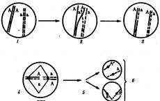
for nucleic acids - Nucleotides. A linear, filamentous molecule of a biopolymer, as a result of the occurrence of hydrogen bonds, has the ability to fit in space in a certain way, for example, in the case of proteins, as shown by L. Pauling, it can take the form of a spiral. This is referred to as a secondary structure. Tertiary structure is said to be when a molecule that has a secondary structure further folds in one way or another, filling three-dimensional space. Finally, molecules that have a three-dimensional structure can enter into interaction, regularly located in space relative to each other and forming what is designated as a quaternary structure; its individual components are commonly referred to as subunits. The most obvious example of how a molecular three-dimensional structure determines the biological functions of a molecule is DNA. It has the structure of a double helix: two threads running in a mutually opposite direction (antiparallel) twist one around the other, forming a double helix with a mutually complementary arrangement of bases, i.e. so that against a certain base of one chain there is always such a the base that best provides the formation of hydrogen bonds: adepine (A) pairs with thymine (T), guanine (G) with cytosine (C). This structure creates optimal conditions for the most important biological functions of DNA: the quantitative multiplication of hereditary information in the process of cell division while maintaining the qualitative invariance of this flow of genetic information. When a cell divides, the strands of the double helix of DNA, which serves as a template, or template, unwind and on each of them, under the action of enzymes, a complementary new strand is synthesized. As a result of this, two completely identical daughter molecules are obtained from one parent DNA molecule (see Cell, Mitosis).
Similarly, in the case of hemoglobin, it turned out that its biological function - the ability to reversibly attach oxygen in the lungs and then give it to tissues - is closely related to the features of the three-dimensional structure of hemoglobin and its changes in the process of implementing its physiological role. When binding and dissociating O 2, spatial changes in the conformation of the hemoglobin molecule occur, leading to a change in the affinity of the iron atoms contained in it for oxygen. Changes in the size of the hemoglobin molecule, reminiscent of changes in the volume of the chest during breathing, made it possible to call hemoglobin "molecular lungs". One of the most important features of living objects is their ability to finely regulate all manifestations of vital activity. M.'s major contribution. scientific discoveries should be considered the discovery of a new, previously unknown regulatory mechanism, referred to as the allosteric effect. It lies in the ability of substances of low molecular weight - the so-called. ligands - modify the specific biological functions of macromolecules, primarily catalytically acting proteins - enzymes, hemoglobin, receptor proteins involved in the construction of biological membranes (See Biological membranes), in synaptic transmission (see Synapses), etc. Three biotic streams. In the light of M.'s ideas. the totality of the phenomena of life can be considered as the result of a combination of three flows: the flow of matter, which finds its expression in the phenomena of metabolism, i.e., assimilation and dissimilation; the flow of energy, which is the driving force for all manifestations of life; and the flow of information that permeates not only the whole variety of processes of development and existence of each organism, but also a continuous series of successive generations. It is the idea of the flow of information, introduced into the doctrine of the living world by the development of biomaterials, that leaves its own specific, unique imprint on it. The most important achievements of molecular biology. Swiftness, scope and depth of M.'s influence. progress in understanding the fundamental problems of studying wildlife is rightly compared, for example, with the influence quantum theory for the development of atomic physics. Two intrinsically related conditions determined this revolutionary impact. On the one hand, a decisive role was played by the discovery of the possibility of studying the most important manifestations of vital activity under the simplest conditions, approaching the type of chemical and physical experiments. On the other hand, as a consequence of this circumstance, there was a rapid involvement of a significant number of representatives of the exact sciences - physicists, chemists, crystallographers, and then mathematicians - in the development of biological problems. Taken together, these circumstances determined the unusually rapid rate of development of M. b., the number and significance of its successes, achieved in just two decades. Here is a far from complete list of these achievements: disclosure of the structure and mechanism of the biological function of DNA, all types of RNA and ribosomes (See Ribosomes) ,
disclosure of the genetic code (See genetic code) ;
discovery of reverse transcription (See transcription) ,
i.e. DNA synthesis on an RNA template; study of the mechanisms of functioning of respiratory pigments; discovery of a three-dimensional structure and its functional role in the action of enzymes (See Enzymes) ,
the principle of matrix synthesis and mechanisms of protein biosynthesis; disclosure of the structure of viruses (See Viruses) and the mechanisms of their replication, the primary and, in part, the spatial structure of antibodies; isolation of individual genes ,
chemical and then biological (enzymatic) gene synthesis, including human, outside the cell (in vitro); transfer of genes from one organism to another, including into human cells; the rapidly progressing deciphering of the chemical structure of an increasing number of individual proteins, mainly enzymes, as well as nucleic acids; detection of the phenomena of "self-assembly" of some biological objects of ever-increasing complexity, starting from nucleic acid molecules and moving on to multicomponent enzymes, viruses, ribosomes, etc.; elucidation of allosteric and other basic principles of regulation of biological functions and processes. Reductionism and integration. M. b. is the final stage of that direction in the study of living objects, which is designated as "reductionism", i.e., the desire to reduce complex life functions to phenomena occurring at the molecular level and therefore accessible to study by the methods of physics and chemistry. Achieved M. b. successes testify to the effectiveness of this approach. At the same time, it must be taken into account that in natural conditions in a cell, tissue, organ, and the whole organism, we are dealing with systems of increasing complexity. Such systems are formed from lower-level components through their regular integration into wholes, acquiring a structural and functional organization and possessing new properties. Therefore, as the knowledge of patterns available for disclosure at the molecular and adjacent levels is detailed, before M. b. the task of understanding the mechanisms of integration as a line further development in the study of life phenomena. The starting point here is the study of the forces of intermolecular interactions - hydrogen bonds, van der Waals, electrostatic forces, etc. By their combination and spatial arrangement, they form what can be designated as "integrative information". It should be considered as one of the main parts of the already mentioned flow of information. In M.'s area. examples of integration can be the phenomena of self-assembly of complex formations from a mixture of their constituent parts. This includes, for example, the formation of multicomponent proteins from their subunits, the formation of viruses from their constituent parts - proteins and nucleic acids, the restoration of the original structure of ribosomes after the separation of their protein and nucleic components, etc. The study of these phenomena is directly related to the knowledge of the main phenomena " recognition” of biopolymer molecules. The point is to find out what combinations of amino acids - in molecules of proteins or nucleotides - in nucleic acids interact with each other during the processes of association of individual molecules with the formation of complexes of a strictly specific, predetermined composition and structure. These include the processes of formation of complex proteins from their subunits; further, selective interaction between nucleic acid molecules, for example, transport and matrix (in this case, the disclosure of the genetic code has significantly expanded our information); finally, this is the formation of many types of structures (for example, ribosomes, viruses, chromosomes), in which both proteins and nucleic acids participate. The disclosure of the corresponding laws, the knowledge of the “language” underlying these interactions, is one of the most important areas of mathematical linguistics, which is still awaiting development. This area is considered as belonging to the number of fundamental problems for the entire biosphere. Problems of molecular biology. Along with the specified important tasks M. would. (knowledge of the patterns of "recognition", self-assembly and integration) an actual direction of scientific search for the near future is the development of methods that allow deciphering the structure, and then the three-dimensional, spatial organization of high-molecular nucleic acids. This has now been achieved with respect to the general plan of the three-dimensional structure of DNA (double helix), but without exact knowledge of its primary structure. Rapid progress in the development of analytical methods allows us to confidently expect the achievement of these goals over the coming years. Here, of course, the main contributions come from representatives of related sciences, primarily physics and chemistry. Everything essential methods, the use of which ensured the emergence and success of M. b., were proposed and developed by physicists (ultracentrifugation, x-ray diffraction analysis, electron microscopy, nuclear magnetic resonance, etc.). Almost all new physical experimental approaches (for example, the use of computers, synchrotron, or bremsstrahlung, radiation, laser technology, and others) open up new opportunities for an in-depth study of the problems of meteorological analysis. Among the most important tasks of a practical nature, the answer to which is expected from M., in the first place is the problem of the molecular basis of malignant growth, then - ways to prevent, and perhaps overcome hereditary diseases - "molecular diseases" (See Molecular diseases ). Of great importance will be the elucidation of the molecular basis of biological catalysis, ie, the action of enzymes. Among the most important modern directions of M. b. should include the desire to decipher the molecular mechanisms of action of hormones (See Hormones) ,
toxic and medicinal substances, as well as to find out the details of the molecular structure and functioning of such cellular structures as biological membranes involved in the regulation of the processes of penetration and transport of substances. More distant goals M. b. - knowledge of the nature of nervous processes, memory mechanisms (See Memory), etc. One of the important emerging sections of M. b. - so-called. genetic engineering, which aims to purposefully operate the genetic apparatus (Genome) of living organisms, starting with microbes and lower (single-celled) and ending with humans (in the latter case, primarily for the purpose of radical treatment of hereditary diseases (See. Hereditary diseases) and the correction of genetic defects ). More extensive interventions in the human genetic basis can only be discussed in a more or less distant future, since in this case serious obstacles, both technical and fundamental, arise. Concerning microbes, plants, and it is possible, and page - x. For animals, such prospects are very encouraging (for example, obtaining varieties of cultivated plants that have an apparatus for fixing nitrogen from the air and do not need fertilizers). They are based on the successes already achieved: the isolation and synthesis of genes, the transfer of genes from one organism to another, the use of mass cell cultures as producers of economic or medically important substances. Organization of research in molecular biology. Fast development M. b. led to the emergence of a large number of specialized research centers. Their number is growing rapidly. The largest: in the UK - the Laboratory of Molecular Biology in Cambridge, the Royal Institute in London; in France - institutes of molecular biology in Paris, Marseille, Strasbourg, the Pasteur Institute; in the USA - departments M. b. at universities and institutes in Boston (Harvard University, Massachusetts Institute of Technology), San Francisco (Berkeley), Los Angeles (California Institute of Technology), New York (Rockefeller University), health institutes in Bethesda, etc.; in Germany - Max Planck institutes, universities in Göttingen and Munich; in Sweden, the Karolinska Institute in Stockholm; in the GDR - the Central Institute for Molecular Biology in Berlin, institutes in Jena and Halle; in Hungary - Biological Center in Szeged. In the USSR the first specialized institute M. would be. was created in Moscow in 1957 in the system of the Academy of Sciences of the USSR (see.
);
then the following were formed: the Institute of Bioorganic Chemistry of the Academy of Sciences of the USSR in Moscow, the Institute of Protein in Pushchino, the Biological Department at the Institute of Atomic Energy (Moscow), and the departments of M. b. at the institutes of the Siberian Branch of the Academy of Sciences in Novosibirsk, the Interdepartmental Laboratory of Bioorganic Chemistry of Moscow State University, the Sector (later Institute) of Molecular Biology and Genetics of the Academy of Sciences of the Ukrainian SSR in Kyiv; significant work on M. b. is conducted at the Institute of Macromolecular Compounds in Leningrad, in a number of departments and laboratories of the Academy of Sciences of the USSR and other departments. Along with individual research centers, organizations of a wider scale arose. In Western Europe, the European Organization for M. arose. (EMBO), in which more than 10 countries participate. In the USSR, in 1966, at the Institute of Molecular Biology, a Scientific Council on M. B. was established, which is the coordinating and organizing center in this field of knowledge. He published an extensive series of monographs on the most important sections of M. b., “winter schools” on M. b. are regularly organized, conferences and symposiums are held on topical problems of M. b. In the future, scientific advice on M. would. were created at the Academy of Medical Sciences of the USSR and many republican Academies of Sciences. The journal Molecular Biology has been published since 1966 (6 issues per year). For rather short term in the USSR the considerable group of researchers in the field of M. has grown; these are scientists of the older generation who have partially switched their interests from other fields; for the most part, they are numerous young researchers. From among the leading scientists who took an active part in the formation and development of M. b. in the USSR, one can name such as A. A. Baev, A. N. Belozersky, A. E. Braunshtein, Yu. A. Ovchinnikov, A. S. Spirin, M. M. Shemyakin, V. A. Engelgardt. M.'s new achievements. and molecular genetics will be facilitated by the resolution of the Central Committee of the CPSU and the Council of Ministers of the USSR (May 1974) "On measures to accelerate the development of molecular biology and molecular genetics and the use of their achievements in the national economy." Lit.: Wagner R., Mitchell G., Genetics and metabolism, trans. from English, M., 1958; Szent-Gyorgy and A., Bioenergetics, trans. from English, M., 1960; Anfinsen K., Molecular basis of evolution, trans. from English, M., 1962; Stanley W., Valens E., Viruses and the nature of life, trans. from English, M., 1963; Molecular genetics, trans. from. English, part 1, M., 1964; Volkenstein M.V., Molecules and life. Introduction to molecular biophysics, M., 1965; Gaurowitz F., Chemistry and functions of proteins, trans. from English, M., 1965; Bresler S. E., Introduction to molecular biology, 3rd ed., M. - L., 1973; Ingram V., Biosynthesis of macromolecules, trans. from English, M., 1966; Engelgardt V. A., Molecular biology, in the book: Development of biology in the USSR, M., 1967; Introduction to molecular biology, trans. from English, M., 1967; Watson, J., Molecular Biology of the Gene, trans. from English, M., 1967; Finean J., Biological ultrastructures, trans. from English, M., 1970; Bendoll, J., Muscles, Molecules, and Movement, trans. from English, M., 1970; Ichas M., Biological code, trans. from English, M., 1971; Molecular biology of viruses, M., 1971; Molecular bases of protein biosynthesis, M., 1971; Bernhard S., Structure and function of enzymes, trans. from English, M., 1971; Spirin A. S., Gavrilova L. P., Ribosome, 2nd ed., M., 1971; Frenkel-Konrat H., Chemistry and biology of viruses, trans. from English, M., 1972; Smith C., Hanewalt F., Molecular Photobiology. Processes of inactivation and recovery, trans. from English, M., 1972; Harris G., Fundamentals of human biochemical genetics, trans. from English, M., 1973. V. A. Engelhardt. Great Soviet Encyclopedia. - M.: Soviet Encyclopedia.
1969-1978
.
The development of biochemistry, biophysics, genetics, cytochemistry, many sections of microbiology and virology around the beginning of the 40s of the XX century. brought close to the study of life phenomena at the molecular level. The successes achieved by these sciences, simultaneously and from different sides, led to the realization of the fact that it is at the molecular level that the main control systems of the body function and that the further progress of these sciences will depend on the disclosure of the biological functions of the molecules that make up the bodies of organisms, their participation in the synthesis and disintegration, mutual transformations and reproduction of compounds in the cell, as well as the exchange of energy and information that occurs in this case. Thus, at the junction of these biological disciplines with chemistry and physics, a completely new branch arose - molecular biology.
Unlike biochemistry, the attention of modern molecular biology is focused mainly on the study of the structure and function of the most important classes of biopolymers - proteins and nucleic acids, the first of which determine the very possibility of metabolic reactions, and the second - the biosynthesis of specific proteins. It is clear, therefore, that it is impossible to make a clear distinction between molecular biology and biochemistry, the corresponding branches of genetics, microbiology, and virology.
The emergence of molecular biology was closely associated with the development of new research methods, which have already been discussed in the relevant chapters. Along with the development of electron microscopy and other methods of microscopic technique, the methods of fractionation of cellular elements developed in the 1950s played an important role. They were based on improved methods of differential centrifugation (A. Claude, 1954). By this time, there were already quite reliable methods for the isolation and fractionation of biopolymers. This includes, in particular, the method of protein fractionation by electrophoresis proposed by A. Tiselius (1937; Nobel Prize, 1948), methods for isolating and purifying nucleic acids (E. Kay, A. Downs, M. Sevag, A. Mirsky, and others. ). At the same time, various methods of chromatographic analysis were developed in many laboratories of the world (A. Martin and R. Sing, 1941; Nobel Prize, 1952), subsequently significantly improved.
X-ray diffraction analysis played an invaluable service in deciphering the structure of biopolymers. The basic principles of X-ray diffraction analysis were developed at King's College London University under the leadership of W. Bragg by a group of researchers, which included J. Bernal, A. Londsdale, W. Astbury, J. Robertson and others.
Of particular note is the research of Professor Moskovsky state university A. R. Kizel on the biochemistry of protoplasm (1925 - 1929), which were of great importance for the subsequent formation of molecular biology. Kizel dealt a blow to the firmly rooted notion that any protoplasm is based on a special protein body - plates, which allegedly determines all its most important structural and functional features. He showed that plates are a protein that is found only in myxomycetes, and then at a certain stage of development, and that no permanent component - a single skeletal protein - exists in protoplasm. Thus, the study of the problem of the structure of protoplasm and the functional role of proteins took the right path and received scope for its development. Kisel's research has won worldwide recognition, stimulating the study of the chemistry of the constituent parts of the cell.
The term "molecular biology", first used by the English crystallographer W. Astbury, professor at the University of Leeds, probably appeared in the early 1940s (before 1945). The fundamental X-ray diffraction studies of proteins and DNA, carried out by Astbury in the 1930s, served as the basis for the subsequent successful deciphering of the secondary structure of these biopolymers. In 1963, J. Bernal wrote: "A monument to him will be erected by the whole of molecular biology - the science that he named and really founded" * , In the literature, this term appeared for the first time, perhaps, in 1946 in the article by W. Astbury "Progress in X-ray diffraction analysis of organic and fibrillar compounds", published in the English journal "Nature" ** . In his Harvey Lecture, Astbury (1950) noted: “I am pleased that the term molecular biology is now quite widely used, although it is unlikely that I was the first to propose it. I liked it and I have long tried to spread it” ***. Already in 1950 Astbury was clear that molecular biology deals primarily with the structure and conformation of macromolecules, the study of which is of decisive importance for understanding the functioning of living organisms.
* (biogr. Mem. Fellows Roy. Soc, 1963, v. 9, 29.)
** (W. T. Astbury. Progress of X-ray analysis of organic and fiber structures.- Nature,. 1946, v. 157, 121.)
*** (W. T. Astbury. Adventures in Molecular Biology. Thomas Springfield, 1952, p. 3.)
Molecular biology has faced and faces, in fact, the same tasks as biology as a whole - the knowledge of the essence of life and its main phenomena, in particular, such as heredity and variability. Modern molecular biology is primarily intended to decipher the structure and function of genes, the ways and mechanisms of realization of the genetic information of organisms at different stages of ontogenesis and at different stages of its reading. It is designed to reveal the subtle mechanisms of regulation of gene activity and cell differentiation, to elucidate the nature of mutagenesis and the molecular basis of the evolutionary process.
Establishing the genetic role of nucleic acids
For the development of molecular biology highest value had the following discoveries. In 1944, American researchers O. Avery, K. McLeod (Nobel Prize, 1923) and M. McCarthy showed that DNA molecules isolated from pneumococci have transforming activity. After hydrolysis of these DNAs by deoxyribonuclease, their transforming activity completely disappeared. Thus, for the first time, it was convincingly proved that it is DNA, and not protein, that is endowed with genetic functions in a cell.
In fairness, it should be noted that the phenomenon of bacterial transformation was discovered much earlier than the discovery of Avery, McLeod and McCarthy. In 1928, F. Griffith published an article in which he reported that after adding killed cells of an encapsulated virulent strain to non-virulent (non-encapsulated) pneumococci, the resulting mixture of cells becomes fatal for mice. Moreover, live pneumococcal cells isolated from animals infected with this mixture were already virulent and possessed a polysaccharide capsule. Thus, in this experiment, it was shown that under the influence of some components of the killed pneumococcal cells, the non-encapsulated form of bacteria turns into a capsule-forming virulent form. Sixteen years later, Avery, McLeod, and McCarthy replaced killed whole pneumococcal cells with their deoxyribonucleic acid in this experiment and showed that it was DNA that had transforming activity (see also chapters 7 and 25). The significance of this discovery is difficult to overestimate. It stimulated the study of nucleic acids in many laboratories around the world and forced scientists to focus on DNA.
Along with the discovery of Avery, McLeod and McCarthy, by the beginning of the 50s, quite a few a large number of direct and indirect evidence that nucleic acids play an exceptional role in life and carry a genetic function. This, in particular, was indicated by the nature of DNA localization in the cell and the data of R. Vendrelli (1948) that the DNA content per cell is strictly constant and correlates with the degree of ploidy: in haploid germ cells, DNA is half that in diploid somatic cells. The pronounced metabolic stability of DNA also testified in favor of the genetic role of DNA. By the beginning of the 50s, a lot of various facts had accumulated, indicating that most of the known mutagenic factors act mainly on nucleic acids and, in particular, on DNA (R. Hotchkiss, 1949; G. Ephrussi-Taylor, 1951; E. Freese , 1957 and others).
Of particular importance in establishing the genetic role of nucleic acids was the study of various phages and viruses. In 1933, D. Schlesinger found DNA in the bacteriophage of Escherichia coli. Since the isolation of tobacco mosaic virus (TMV) in the crystalline state by W. Stanley (1935, Nobel Prize, 1946), a new stage in the study of plant viruses has begun. In 1937 - 1938. employees of the Rothamsted Agricultural Station (England) F. Bowden and N. Pirie showed that many plant viruses isolated by them are not globulins, but are ribonucleoproteins and contain nucleic acid as an obligatory component. At the very beginning of the 40s, the works of G. Schramm (1940), P. A. Agatov (1941), G. Miller and W. Stanley (1941) were published, indicating that a noticeable chemical modification of the protein component does not lead to loss of TMV infectivity. This indicated that the protein component could not be the carrier of the hereditary properties of the virus, as many microbiologists continued to believe. Convincing evidence in favor of the genetic role of nucleic acid (RNA) in plant viruses was obtained in 1956 by G. Schramm in Tübingen (FRG) and H. Frenkel-Konrath in California (USA). These researchers almost simultaneously and independently of each other isolated RNA from TMV and showed that it, and not the protein, has infectivity: as a result of infection of tobacco plants with this RNA, normal viral particles were formed and multiplied in them. This meant that RNA contained information for the synthesis and assembly of all viral components, including the viral protein. In 1968, I. G. Atabekov established that protein plays a significant role in the very infection of plants - the nature of the protein determines the spectrum of host plants.
In 1957, Frenkel-Konrat for the first time carried out the reconstruction of the TMV from its constituent components - RNA and protein. Along with normal particles, he received mixed "hybrids" in which the RNA was from one strain and the protein from another. The heredity of such hybrids was completely determined by RNA, and the progeny of the viruses belonged to the strain whose RNA was used to obtain the initial mixed particles. Later, the experiments of A. Gierer, G. Schuster and G. Schramm (1958) and G. Witman (1960 - 1966) showed that the chemical modification of the TMV nucleic component leads to the appearance of various mutants of this virus.
In 1970, D. Baltimore and G. Temin found that the transfer of genetic information can occur not only from DNA to RNA, but vice versa. They found in some oncogenic RNA-containing viruses (oncornaviruses) a special enzyme, the so-called reverse transcriptase, which is capable of synthesizing complementary DNA on RNA chains. This major discovery made it possible to understand the mechanism of insertion of the genetic information of RNA-containing viruses into the host genome and to take a fresh look at the nature of their oncogenic action.
Discovery of nucleic acids and study of their properties
The term nucleic acids was introduced by the German biochemist R. Altman in 1889, after these compounds were discovered in 1869 by the Swiss physician F. Miescher. Misher extracted the pus cells with dilute hydrochloric acid for several weeks and obtained almost pure nuclear material in the remainder. He considered this material to be a characteristic "substance of cell nuclei and called it nuclein. In terms of its properties, nuclein differed sharply from proteins: it was more acidic, did not contain sulfur, but it contained a lot of phosphorus, it was readily soluble in alkalis, but did not dissolve in dilute acids.
Misher sent the results of his observations on nuclein to F. Goppe-Seyler for publication in a journal. The substance he described was so unusual (at that time only lecithin was known of all biological phosphorus-containing compounds) that Goppe-Seyler did not believe Misher's experiments, returned the manuscript to him and instructed his employees N. Plosh and N. Lyubavin to check his conclusions on other material . Miescher's work "On the chemical composition of pus cells" was published two years later (1871). At the same time, the works of Goppe-Seyler and his collaborators were published on the composition of pus cells, erythrocytes of birds, snakes, and other cells. Over the next three years, nuclein was isolated from animal cells and yeast.
In his work, Misher noted that a detailed study of different nucleins can lead to the establishment of differences between them, thereby anticipating the idea of specificity of nucleic acids. While studying salmon milk, Misher found that the nuclein in them is in the form of salt and is associated with the main protein, which he called protamine.
In 1879, A. Kossel began to study nucleins in the laboratory of Goppe-Seyler. In 1881, he isolated hypoxanthine from nuclein, but at that time he still doubted the origin of this base and believed that hypoxanthine could be a degradation product of proteins. In 1891, among the products of nuclein hydrolysis, Kossel discovered adenine, guanine, phosphoric acid, and another substance with the properties of sugar. For research on the chemistry of nucleic acids, Kossel was awarded the Nobel Prize in 1910.
Further progress in deciphering the structure of nucleic acids is associated with the research of P. Levin and colleagues (1911 - 1934). In 1911, P. Levin and V. Jacobs identified the carbohydrate component of adenosine and guanosine; they found that these nucleosides contain D-ribose. In 1930, Lewin showed that the carbohydrate component of deoxyribonucleosides is 2-deoxy-D-ribose. From his work, it became known that nucleic acids are built from nucleotides, i.e., phosphorylated nucleosides. Levin believed that the main type of bond in nucleic acids (RNA) is the 2", 5" phosphodiester bond. This notion turned out to be wrong. Thanks to the work of the English chemist A. Todd (Nobel Prize, 1957) and his collaborators, as well as the English biochemists R. Markham and J. Smith, it became known in the early 50s that the main type of bond in RNA is 3", 5" - phosphodiester bond.
Lewin showed that different nucleic acids can differ in the nature of the carbohydrate component: some of them contain the sugar deoxyribose, while others contain ribose. In addition, these two types of nucleic acids differed in the nature of one of the bases: pentose-type nucleic acids contained uracil, and deoxypentose-type nucleic acids contained thymine. Deoxypentose nucleic acid (in modern terminology, deoxyribonucleic acid - DNA) was usually easily isolated in large quantities from the thymus (sweet gland) of calves. Therefore, it was called thymonucleic acid. The source of pentose-type nucleic acid (RNA) was mainly yeast and wheat germ. This type was often referred to as yeast nucleic acid.
In the early 1930s, the notion that plant cells were characterized by a yeast-type nucleic acid was rather firmly rooted, while thymonucleic acid was characteristic only of the nuclei of animal cells. The two types of nucleic acids, RNA and DNA, were then called plant and animal nucleic acids, respectively. However, as the early studies of A. N. Belozersky showed, such a division of nucleic acids is unjustified. In 1934, Belozersky first discovered thymonucleic acid in plant cells: from pea seedlings, he isolated and identified the thymine-pyrimidine base, which is characteristic of DNA. Then he discovered thymine in other plants (soybean seeds, beans). In 1936, A. N. Belozersky and I. I. Dubrovskaya isolated DNA preparatively from horse chestnut seedlings. In addition, a series of studies carried out in England in the 1940s by D. Davidson and co-workers convincingly showed that plant nucleic acid (RNA) is contained in many animal cells.
The widespread use of the cytochemical reaction for DNA developed by R. Felgen and G. Rosenbeck (1924) and the reaction of J. Brachet (1944) for RNA made it possible to quickly and unambiguously resolve the issue of the preferential localization of these nucleic acids in the cell. It turned out that DNA is concentrated in the nucleus, while RNA is predominantly concentrated in the cytoplasm. Later, it was found that RNA is contained both in the cytoplasm and in the nucleus, and in addition, cytoplasmic DNA was identified.
As for the question of the primary structure of nucleic acids, by the mid-1940s, P. Levin's idea was firmly established in science, according to which all nucleic acids are built according to the same type and consist of the same so-called tetranucleotide blocks. Each of these blocks, according to Lewin, contains four different nucleotides. The tetranucleotide theory of the structure of nucleic acids largely deprived these biopolymers of specificity. Therefore, it is not surprising that at that time all the specifics of living things were associated only with proteins, the nature of the monomers of which is much more diverse (20 amino acids).
The first gap in the theory of the tetranucleotide structure of nucleic acids was made by the analytical data of the English chemist J. Gouland (1945 - 1947). When determining the composition of nucleic acids by the base nitrogen, he did not obtain an equimolar ratio of bases, as it should have been according to Lewin's theory. Finally, the tetranucleotide theory of the structure of nucleic acids collapsed as a result of the research of E. Chargaff and his collaborators (1949 - 1951). Chargaff used paper chromatography to separate the bases released from DNA as a result of its acid hydrolysis. Each of these bases was accurately determined spectrophotometrically. Chargaff noticed significant deviations from the equimolar ratio of bases in DNA of different origins and for the first time definitely stated that DNA has a pronounced species specificity. This ended the hegemony of the concept of protein specificity in the living cell. Analyzing DNA of different origins, Chargaff discovered and formulated unique patterns of DNA composition, which entered science under the name of Chargaff's rules. According to these rules, in all DNA, regardless of origin, the amount of adenine is equal to the amount of thymine (A = T), the amount of guanine is equal to the amount of cytosine (G = C), the amount of purines is equal to the amount of pyrimidines (G + A = C + T), the amount bases with 6-amino groups is equal to the number of bases with 6-keto groups (A + C = G + T). At the same time, despite such strict quantitative correspondences, DNA of different species differ in the value of the A+T:G+C ratio. In some DNA, the amount of guanine and cytosine prevails over the amount of adenine and thymine (Chargaff called these DNA GC-type DNA); other DNAs contained more adenine and thymine than guanine and cytosine (these DNAs were called AT-type DNA). The data obtained by Chargaff on the composition of DNA played an exceptional role in molecular biology. It was they that formed the basis for the discovery of the structure of DNA, made in 1953 by J. Watson and F. Crick.
Back in 1938, W. Astbury and F. Bell, using X-ray diffraction analysis, showed that the base planes in DNA should be perpendicular to the long axis of the molecule and resemble, as it were, a stack of plates lying one above the other. With the improvement of the technique of X-ray diffraction analysis, by 1952 - 1953. accumulated information that made it possible to judge the length of individual bonds and the angles of inclination. This made it possible to represent with the greatest probability the nature of the orientation of the rings of pentose residues in the sugar-phosphate backbone of the DNA molecule. In 1952, S. Farberg proposed two speculative models of DNA, which represented a single-stranded molecule folded or twisted on itself. A no less speculative model of the structure of DNA was proposed in 1953 by L. Pauling (Nobel Prize winner, 1954) and R. Corey. In this model, three twisted strands of DNA formed a long helix, the core of which was represented by phosphate groups, and the bases were located outside of it. By 1953, M. Wilkins and R. Franklin obtained clearer X-ray diffraction patterns of DNA. Their analysis showed the complete failure of the models of Farberg, Pauling and Corey. Using Chargaff's data, comparing different combinations of molecular models of individual monomers and X-ray diffraction data, J. Watson and F. Crick in 1953 came to the conclusion that the DNA molecule must be a double-stranded helix. Chargaff's rules severely limited the number of possible ordered combinations of bases in the proposed DNA model; they suggested to Watson and Crick that there must be a specific base pairing in the DNA molecule - adenine with thymine, and guanine with cytosine. In other words, adenine in one strand of DNA always strictly corresponds to thymine in the other strand, and guanine in one strand necessarily corresponds to cytosine in the other. Thus Watson and Crick for the first time formulated the principle of the complementary structure of DNA of exceptional importance, according to which one strand of DNA complements another, i.e., the base sequence of one strand uniquely determines the sequence of bases in the other (complementary) strand. It became obvious that already in the very structure of DNA lies the potential for its exact reproduction. This model of DNA structure is currently generally accepted. Crick, Watson and Wilkins were awarded the Nobel Prize in 1962 for deciphering the structure of DNA.
It should be noted that the idea of a mechanism for the exact reproduction of macromolecules and the transmission of hereditary information originated in our country. In 1927, N. K. Koltsov suggested that during cell reproduction, the reproduction of molecules occurs by exact autocatalytic reproduction of the existing parent molecules. True, at that time Koltsov endowed this property not with DNA molecules, but with molecules of a protein nature, the functional significance of which was then unknown. Nevertheless, the very idea of autocatalytic reproduction of macromolecules and the mechanism of transmission of hereditary properties turned out to be prophetic: it became the guiding idea of modern molecular biology.
Conducted in the laboratory of A. N. Belozersky by A. S. Spirin, G. N. Zaitseva, B. F. Vanyushin, S. O. Uryson, A. S. Antonov and others variety of organisms fully confirmed the patterns discovered by Chargaff, and full compliance with the molecular model of the structure of DNA proposed by Watson and Crick. These studies have shown that the DNA of different bacteria, fungi, algae, actinomycetes, higher plants, invertebrates and vertebrates have a specific composition. Differences in the composition (the content of AT-base pairs) are especially pronounced in microorganisms, turning out to be an important taxonomic feature. In higher plants and animals, species variations in the composition of DNA are much less pronounced. But this does not mean that their DNA is less specific. In addition to the composition of bases, specificity is largely determined by their sequence in DNA chains.
Along with the usual bases, additional nitrogenous bases were found in DNA and RNA. Thus, G. White (1950) found 5-methylcytosine in the DNA of plants and animals, and D. Dunn and J. Smith (1958) found methylated adenine in some DNA. For a long time, methylcytosine was considered a hallmark of the genetic material of higher organisms. In 1968, A. N. Belozersky, B. F. Vanyushin and N. A. Kokurina found that it can also be found in the DNA of bacteria.
In 1964, M. Gold and J. Hurwitz discovered a new class of enzymes that carry out the natural modification of DNA - its methylation. After this discovery, it became clear that minor (contained in small amounts) bases arise already on the finished DNA polynucleotide chain as a result of specific methylation of cytosine and adenine residues in special sequences. In particular, according to B. F. Vanyushin, Ya. I. Buryanov, and A. N. Belozersky (1969), adenine methylation in E. coli DNA can occur in terminating codons. According to A. N. Belozersky and colleagues (1968 - 1970), as well as M. Meselson (USA) and V. Arber (Switzerland) (1965 - 1969), methylation gives unique individual features to DNA molecules and, in combination with the action of specific nucleases, is part of a complex mechanism that controls the synthesis of DNA in the cell. In other words, the nature of methylation of a particular DNA predetermines the question of whether it can multiply in a given cell.
Almost at the same time, the isolation and intensive study of DNA methylases and restriction endonucleases began; in 1969 - 1975 the nucleotide sequences recognized in DNA by some of these enzymes have been established (X. Boyer, X. Smith, S. Lynn, K. Murray). When different DNAs are hydrolyzed by a restriction enzyme, rather large fragments with identical "sticky" ends are cleaved out. This makes it possible not only to analyze the structure of genes, as is done in small viruses (D. Nathans, S. Adler, 1973 - 1975), but also to construct various genomes. With the discovery of these specific restriction enzymes, genetic engineering has become a tangible reality. Embedded in small plasmid DNA genes of various origins are already easily introduced into various cells. So, a new type of biologically active plasmids was obtained, giving resistance to certain antibiotics (S. Cohen, 1973), ribosomal genes of a frog and Drosophila were introduced into Escherichia coli plasmids (J. Morrow, 1974; X. Boyer, D. Hogness, R. Davis , 1974 - 1975). Thus, real ways are open for obtaining fundamentally new organisms by introducing and integrating various genes into their gene pool. This discovery can be directed to the benefit of all mankind.
In 1952, G. White and S. Cohen discovered that the DNA of T-even phages contains an unusual base - 5-hydroxymethylcytosine. Later, from the works of E. Volkin and R. Sinsheimer (1954) and Cohen (1956), it became known that hydroxymethylcytosine residues can be completely or partially glucosidized, as a result of which the phage DNA molecule is protected from the hydrolytic action of nucleases.
In the early 1950s, from the works of D. Dunn and J. Smith (England), S. Zamenhof (USA) and A. Wacker (Germany), it became known that many artificial base analogues can be included in DNA, sometimes replacing up to 50% thymine. As a rule, these substitutions lead to errors in DNA replication, transcription and translation and to the appearance of mutants. Thus, J. Marmur (1962) found that the DNA of some phages contains oxymethyluracil instead of thymine. In 1963, I. Takahashi and J. Marmur discovered that the DNA of one of the phages contains uracil instead of thymine. Thus, another principle, according to which nucleic acids were previously separated, collapsed. Since the time of P. Levin's work, it has been believed that thymine is the hallmark of DNA, and uracil is the hallmark of RNA. It became clear that this sign is not always reliable, and the fundamental difference in the chemical nature of the two types of nucleic acids, as it seems today, is only the nature of the carbohydrate component.
In the study of phages, many unusual features of the organization of nucleic acids have been uncovered. Since 1953, it has been believed that all DNA are double-stranded linear molecules, while RNA is only single-stranded. This position was significantly shaken in 1961, when R. Sinsheimer discovered that the DNA of the phage φ X 174 is represented by a single-stranded circular molecule. However, later it turned out that in this form this DNA exists only in a vegetative phage particle, and the replicative form of the DNA of this phage is also double-stranded. In addition, it turned out to be quite unexpected that the RNA of some viruses can be double-stranded. This new type of macromolecular organization of RNA was discovered in 1962 by P. Gomatos, I. Tamm and other researchers in some animal viruses and in plant wound tumor virus. Recently, V. I. Agol and A. A. Bogdanov (1970) established that in addition to linear RNA molecules, there are also closed or cyclic molecules. They detected cyclic double-stranded RNA, in particular, in the encephalomyelocarditis virus. Thanks to the works of X. Deveaux, L. Tinoko, T. I. Tikhonenko, E. I. Budovsky and others (1960 - 1974), the main features of the organization (laying) of genetic material in bacteriophages became known.
In the late 1950s, the American scientist P. Doty found that heating causes DNA denaturation, which is accompanied by the breaking of hydrogen bonds between base pairs and the separation of complementary chains. This process has the character of a "spiral-coil" phase transition and resembles the melting of crystals. Therefore, Doty called the process of thermal denaturation of DNA DNA melting. With slow cooling, renaturation of the molecules occurs, i.e., the reunification of the complementary halves.
The principle of renaturation in 1960 was used by J. Marmur and K. Schildkraut to determine the degree of "hybridization" of DNA of different microorganisms. Subsequently, E. Bolton and B. McCarthy improved this technique by proposing the method of the so-called DNA-agar columns. This method turned out to be indispensable in studying the degree of homology of the nucleotide sequence of different DNA and elucidating the genetic relationship of different organisms. The denaturation of DNA discovered by Doty in combination with the chromatography on methylated albumin described by J. Mandel and A. Hershey * (1960) and density gradient centrifugation (the method was developed in 1957 by M. Meselson, F. Stahl and D. Winograd) is widely used for separation, isolation and analysis of individual complementary DNA strands For example, W. Shibalsky (USA), using these techniques to separate the DNA of the lambda phage, showed in 1967 - 1969 that both phage chains are genetically active, and not one, as this was considered to be (S. Spiegelman, 1961). It should be noted that for the first time the idea of the genetic significance of both DNA strands of the lambda phage was expressed in the USSR by SE Bresler (1961).
* (For their work on the genetics of bacteria and viruses, A. Hershey, together with M. Delbrück and S. Luria, were awarded the Nobel Prize in 1969.)
To understand the organization and functional activity of the genome, the determination of the DNA nucleotide sequence is of paramount importance. The search for methods for such determination is carried out in many laboratories around the world. Since the late 1950s, M. Beer and his collaborators have been trying to establish the DNA sequence using electron microscopy in the USA, but so far without success. In the early 1950s, from the first works of Sinsheimer, Chargaff, and other researchers on the enzymatic degradation of DNA, it became known that different nucleotides in a DNA molecule are distributed, although not randomly, but unevenly. According to the English chemist C. Barton (1961), pyrimidines (more than 70%) are concentrated mainly in the form of the corresponding blocks. A. L. Mazin and B. F. Vanyushin (1968 - 1969) found that different DNAs have different degrees of pyrimidine cohesion and that in the DNA of animal organisms it increases markedly as it moves from lower to higher. Thus, the evolution of organisms is also reflected in the structure of their genomes. That is why, for understanding the evolutionary process as a whole, the comparative study of the structure of nucleic acids is of particular importance. Analysis of the structure of biologically important polymers and, first of all, DNA is extremely important for solving many particular problems of phylogenetics and taxonomy.
It is interesting to note that the English physiologist E. Lankester, who studied the hemoglobins of mollusks, anticipated the ideas of molecular biology exactly 100 years ago, wrote: “Chemical differences between different species and genera of animals and plants are as important for clarifying the history of their origin as their form. If we could clearly establish the differences in the molecular organization and functioning of organisms, we would be able to understand the origin and evolution of different organisms much better than on the basis of morphological observations " * . The significance of biochemical studies for taxonomy was also emphasized by V. L. Komarov, who wrote that “the basis of all even purely morphological characters, on the basis of which we classify and establish species, are precisely biochemical differences” **.
* (E. R. Lankester. Uber das Vorcommen von Haemoglobin in den Muskeln der Mollusken und die Verbreitung desselben in den lebendigen Organismen.- "Pfluger" s Archiv fur die gesammte Physiol., 1871, Bd 4, 319.)
** (V. L. Komarov. Selected works, vol. 1. M.-L., Publishing House of the Academy of Sciences of the USSR, 1945, p. 331.)
A. V. Blagoveshchenskii and S. L. Ivanov, back in the 1920s, took the first steps in our country to elucidate certain questions of the evolution and systematics of organisms on the basis of a comparative analysis of their biochemical composition (see Chapter 2). Comparative analysis the structure of proteins and nucleic acids is now becoming an increasingly tangible tool for taxonomists (see Chapter 21). This method of molecular biology makes it possible not only to clarify the position of individual species in the system, but also makes it necessary to take a fresh look at the very principles of classification of organisms, and sometimes to revise the entire system as a whole, as happened, for example, with the systematics of microorganisms. Undoubtedly, in the future, the analysis of the structure of the genome will take central location in the chemosystematics of organisms.
Of great importance for the development of molecular biology was the deciphering of the mechanisms of DNA replication and transcription (see Chapter 24).
Protein biosynthesis
An important shift in solving the problem of protein biosynthesis is associated with advances in the study of nucleic acids. In 1941, T. Kasperson (Sweden) and in 1942, J. Brachet (Belgium) drew attention to the fact that tissues with active protein synthesis contain an increased amount of RNA. They concluded that ribonucleic acids play a decisive role in protein synthesis. In 1953, E. Gale and D. Fox seem to have received direct evidence of the direct involvement of RNA in protein biosynthesis: according to their data, ribonuclease significantly suppressed the incorporation of amino acids in bacterial cell lysates. Similar data were obtained by V. Olfri, M. Delhi and A. Mirsky (1953) on liver homogenates. Later, E. Gale rejected his correct idea of the leading role of RNA in protein synthesis, mistakenly believing that the activation of protein synthesis in a cell-free system occurred under the influence of some other substance of an unknown nature. In 1954, P. Zamechnik, D. Littlefield, R. B. Khesin-Lurie and others found that the most active incorporation of amino acids occurs in RNA-rich fractions of subcellular particles - microsomes. P. Zamechnik and E. Keller (1953 - 1954) found that the incorporation of amino acids was noticeably enhanced in the presence of the supernatant under conditions of ATP regeneration. P. Sikevitz (1952) and M. Hoagland (1956) isolated a protein fraction (pH 5 fraction) from the supernatant, which was responsible for the sharp stimulation of the inclusion of amino acids in microsomes. Along with proteins, a special class of low molecular weight RNAs, now called transfer RNAs (tRNAs), was found in the supernatant. In 1958, Hoagland and Zamechnik, as well as P. Berg, R. Sweet and F. Allen and many other researchers found that each amino acid requires its own special enzyme, ATP and specific tRNA, to activate. It became clear that tRNAs perform exclusively the function of adapters, i.e. devices that find a place on the nucleic matrix (mRNA) for the corresponding amino acid in the emerging protein molecule. These studies fully confirmed the adapter hypothesis of F. Crick (1957), which provided for the existence of polynucleotide adapters in the cell, which are necessary for the correct arrangement of amino acid residues of the synthesized protein on the nucleic matrix. Much later, the French scientist F. Chapville (1962) in the laboratory of F. Lipman (Nobel Prize, 1953) in the USA very ingeniously and unequivocally showed that the location of an amino acid in a synthesized protein molecule is completely determined by the specific tRNA to which it is attached. Crick's adaptor hypothesis was developed by Hoagland and Zamechnik.
By 1958, the following main stages of protein synthesis became known: 1) activation of an amino acid by a specific enzyme from the "pH 5 fraction" in the presence of ATP with the formation of aminoacyl adenylate; 2) attachment of an activated amino acid to a specific tRNA with the release of adenosine monophosphate (AMP); 3) binding of aminoacyl-tRNA (tRNA loaded with an amino acid) to microsomes and incorporation of amino acids into a protein with tRNA release. Hoagland (1958) noted that guanosine triphosphate (GTP) is required at the last stage of protein synthesis.
Transfer RNAs and gene synthesis
After the discovery of tRNAs, active searches for their fractionation and determination of the nucleotide sequence began. The American biochemist R. Holly achieved the greatest success. In 1965, he established the structure of alanine tRNA from yeast. Using ribonucleases (guanyl RNase and pancreatic RNase), Holly divided the nucleic acid molecule into several fragments, determined the nucleotide sequence in each of them separately, and then reconstructed the sequence of the entire alanine tRNA molecule. This way of analyzing the nucleotide sequence is called the block method. Holly's merit consisted mainly in the fact that he learned to divide the RNA molecule not only into small pieces, as many did before him, but also into large fragments (quarters and halves). This gave him the opportunity to correctly assemble individual small pieces together and thereby recreate the complete nucleotide sequence of the entire tRNA molecule (Nobel Prize, 1968).
This technique was immediately adopted by many laboratories around the world. Over the next two years, the primary structure of several tRNAs was deciphered in the USSR and abroad. A. A. Baev (1967) and co-workers established the nucleotide sequence in yeast valine tRNA for the first time. To date, more than a dozen different individual tRNAs have been studied. A peculiar record in determining the nucleotide sequence was set in Cambridge by F. Senger and G. Brownlee. These researchers developed a surprisingly elegant method for separating oligonucleotides and sequencing the so-called 5 S (ribosomal) RNA from E. coli cells (1968). This RNA consists of 120 nucleotide residues and, unlike tRNA, does not contain additional minor bases, which greatly facilitate the analysis of the nucleotide sequence, serving as unique landmarks for individual fragments of the molecule. At present, thanks to the use of the method of Sanger and Brownlee, work on the study of the sequence of long ribosomal RNAs and some viral RNAs is being successfully advanced in the laboratory of J. Ebel (France) and other researchers.
A. A. Baev and colleagues (1967) found that valine tRNA cut in half restores its macromolecular structure in solution and, despite a defect in the primary structure, has the functional activity of the original (native) molecule. This approach - the reconstruction of a cut macromolecule after the removal of certain fragments - turned out to be very promising. It is now widely used to elucidate the functional role of individual sections of certain tRNAs.
In recent years, great success has been achieved in obtaining crystalline preparations of individual tRNAs. Many tRNAs have already been crystallized in several laboratories in the USA and England. This made it possible to study the structure of tRNA using X-ray diffraction analysis. In 1970, R. Bock presented the first X-ray patterns and three-dimensional models of several tRNAs that he had created at the University of Wisconsin. These models help determine the localization of individual functionally active sites in tRNA and understand the basic principles of the functioning of these molecules.
The deciphering of the nature of the genetic code (see Chapter 24), which, without exaggeration, can be regarded as the leading achievement of natural science in the 20th century, was of paramount importance for revealing the mechanism of protein synthesis and solving the problem of the specificity of this process.
R. Holly's discovery of the primary structure of tRNA gave impetus to the work of G. Korana * (USA) on the synthesis of oligonucleotides and directed them towards the synthesis of a specific biological structure - a DNA molecule encoding alanine tRNA. The first steps in the chemical synthesis of short oligonucleotides made by the Qur'an almost 15 years ago culminated in 1970 with the first gene synthesis. Koran and his collaborators first chemically synthesized short fragments of 8-12 nucleotide residues from individual nucleotides. These fragments with a given nucleotide sequence formed spontaneously double-stranded complementary pieces with an overlap of 4–5 nucleotides. Then these ready-made pieces were joined end-to-end in the right order using the enzyme DNA ligase. Thus, in contrast to the replication of DNA molecules, according to A. Kornberg ** (see Chapter 24), the Qur'an managed to re-create a natural double-stranded DNA molecule according to a pre-planned program in accordance with the tRNA sequence described by Holly. Similarly, work is now underway on the synthesis of other genes (M. N. Kolosov, Z. A. Shabarova, D. G. Knorre, 1970 - 1975).
* (For the study of the genetic code, G. Koran and M. Nirenberg were awarded the Nobel Prize in 1968.)
** (For the discovery of polymerase and DNA synthesis A. Kornberg, and for the synthesis of RNA S. Ochoa in 1959 was awarded the Nobel Prize.)
Microsomes, ribosomes, translation
In the mid-1950s, it was believed that microsomes were the center of protein synthesis in the cell. The term microsomes was first introduced in 1949 by A. Claude to refer to the fraction of small granules. Later it turned out that not the entire fraction of microsomes, consisting of membranes and granules, but only small ribonucleoprotein particles, is responsible for protein synthesis. These particles in 1958 were called ribosomes by R. Roberts.
Classical studies of bacterial ribosomes were carried out by A. Tisier and J. Watson in 1958-1959. Bacterial ribosomes turned out to be somewhat smaller than plant and animal ones. J. Littleton (1960), M. Clark (1964) and E. N. Svetailo (1966) showed that the ribosomes of the chloroplasts of higher plants and mitochondria belong to the bacterial type. A. Tisier and others (1958) found that ribosomes dissociate into two unequal subunits containing one RNA molecule each. In the late 50s, it was believed that each ribosomal RNA molecule consists of several short fragments. However, AS Spirin in 1960 was the first to show that RNA in subparticles is represented by a continuous molecule. D. Waller (1960), having separated ribosomal proteins using starch gel electrophoresis, found that they are very heterogeneous. At first, many doubted Waller's data, since it seemed that the ribosome protein should be strictly homogeneous, like, for example, the TMV protein. At present, as a result of the research of D. Waller, R. Trout, P. Traub and other biochemists, it has become known that the composition of the actual ribosomal particles includes more than 50 proteins that are completely different in structure. AS Spirin in 1963 was the first to unfold ribosomal subparticles and show that ribosomes are a compactly twisted ribonucleoprotein strand, which can unfold under certain conditions. In 1967 - 1968 M. Nomura completely reconstructed a biologically active subunit from ribosomal RNA and protein and even obtained ribosomes in which protein and RNA belonged to different microorganisms.
The role of ribosomal RNA is still unclear. It is assumed that it is that unique specific matrix on which, during the formation of a ribosomal particle, each of the numerous ribosomal proteins finds a strictly defined place (AS Spirin, 1968).
A. Rich (1962) discovered aggregates of several ribosomes interconnected by a strand of mRNA. These complexes were called polysomes. The discovery of polysomes allowed Rich and Watson (1963) to suggest that the synthesis of the polypeptide chain occurs on the ribosome, which, as it were, moves along the mRNA chain. As the ribosome moves along the mRNA chain, information is read out in the particle and the protein polypeptide chain is formed, and new ribosomes alternately attach to the released read end of the mRNA. From the data of Rich and Watson, it followed that the significance of polysomes in a cell lies in the mass production of protein by successive reading of the matrix by several ribosomes at once.
As a result of the research of M. Nirenberg, S. Ochoa, F. Lipman, G. Korana and others in 1963 - 1970. it became known that along with mRNA, ribosomes, ATP and aminoacyl-tRNA, a large number of various factors take part in the translation process, and the translation process itself can be conditionally divided into three stages - initiation, translation itself and termination.
Translation initiation means the synthesis of the first peptide bond in the complex ribosome - template polynucleotide - aminoacyl-tRNA. Such initiatory activity is possessed not by any aminoacyl-tRNA, but by formylmethionyl-tRNA. This substance was first isolated in 1964 by F. Senger and K. Marker. S. Bretcher and K. Marker (1966) showed that the initiatory function of formylmethionyl-tRNA is due to its increased affinity for the peptidyl center of the ribosome. For the start of translation, some protein initiation factors are also extremely important, which were isolated in the laboratories of S. Ochoa, F. Gro and other research centers. After the formation of the first peptide bond in the ribosome, translation itself begins, i.e., the sequential addition of an aminoacyl residue to the C-terminus of the polypeptide. Many details of the translation process were studied by K. Monroe and J. Bishop (England), I. Rykhlik and F. Shorm (Czechoslovakia), F. Lipman, M. Bretcher, W. Gilbert (USA) and other researchers. In 1968, A. S. Spirin proposed an original hypothesis to explain the mechanism of the ribosome. The driving mechanism that ensures all spatial movements of tRNA and mRNA during translation is the periodic opening and closing of ribosome subparticles. The translation termination is encoded in the readable matrix itself, which contains the termination codons. As shown by S. Brenner (1965 - 1967), triplets UAA, UAG and UGA are such codons. M. Capecci (1967) also identified special protein termination factors. AS Spirin and LP Gavrilova described the so-called "non-enzymatic" protein synthesis in ribosomes (1972 - 1975) without the participation of protein factors. This discovery is important for understanding the origin and evolution of protein biosynthesis.
Regulation of gene and protein activity
After the problem of the specificity of protein synthesis, the problem of regulation of protein synthesis, or, what is the same, regulation of gene activity, turned out to be in the first place in molecular biology.
The functional inequivalence of cells and the repression and activation of genes associated with it have long attracted the attention of geneticists, but until recently the real mechanism for controlling gene activity remained unknown.
The first attempts to explain the regulatory activity of genes were associated with the study of histone proteins. Even the Steadman spouses * in the early 40s of the XX century. suggested that it is histones that can play the main role in this phenomenon. Subsequently, they obtained the first clear data on differences in the chemical nature of histone proteins. At present, the number of facts testifying in favor of this hypothesis is increasing every year.
* (E. Stedman, E. Stedman. The basic proteins of cell nuclei.- Philosoph. Trans. Roy. soc. London, 1951, v. 235, 565 - 595.)
At the same time, an increasing amount of data is accumulating, indicating that the regulation of gene activity is a much more complex process than the simple interaction of gene sections with histone protein molecules. In 1960 - 1962 in the laboratory of R. B. Khesin-Lurie, it was found that phage genes begin to be read non-simultaneously: T2 phage genes can be divided into early ones, the functioning of which occurred in the first minutes of infection of a bacterial cell, and late ones, which began to synthesize mRNA after the completion of the work of early genes.
In 1961, the French biochemists F. Jacob and J. Monod proposed a scheme for the regulation of gene activity, which played an exceptional role in understanding the regulatory mechanisms of the cell in general. According to the scheme of Jacob and Monod, in addition to structural (informational) genes, DNA also contains genes-regulators and genes-operators. The regulator gene encodes the synthesis of a specific substance - a repressor, which can attach both to the inducer and to the operator gene. The operator gene is linked to structural genes, while the regulator gene is located at some distance from them. If there is no inducer in the environment, for example, lactose, then the repressor synthesized by the regulator gene binds to the operator gene and, blocking it, turns off the work of the entire operon (a block of structural genes together with the operator that controls them). Enzyme formation does not occur under these conditions. If an inductor (lactose) appears in the medium, then the product of the regulator gene, the repressor, binds to lactose and removes the block from the operator gene. In this case, the work of the structural gene encoding the synthesis of the enzyme becomes possible, and the enzyme (lactose) appears in the medium.
According to Jacob and Monod, this regulation scheme is applicable to all adaptive enzymes and can take place both during repression, when the formation of the enzyme is suppressed by an excess of the reaction product, and during induction, when the introduction of a substrate causes the synthesis of the enzyme. For studies of the regulation of gene activity, Jacob and Monod were awarded the Nobel Prize in 1965.
Initially, this scheme seemed too far-fetched. However, later it turned out that the regulation of genes according to this principle takes place not only in bacteria, but also in other organisms.
Since 1960, a prominent place in molecular biology has been occupied by studies of the organization of the genome and the structure of chromatin in eukaryotic organisms (J. Bonner, R. Britten, W. Olfrey, P. Walker, Yu. S. Chentsov, I. B. Zbarsky and others .) and regulation of transcription (A. Mirsky, G. P. Georgiev, M. Bernstiel, D. Goll, R. Tsanev, R. I. Salganik). For a long time, the nature of the repressor remained unknown and controversial. In 1968, M. Ptashne (USA) showed that a protein is a repressor. He isolated it in the laboratory of J. Watson and found that the repressor indeed has an affinity for the inductor (lactose) and at the same time "recognizes" the operator gene of the lac operon and specifically binds to it.
In the last 5 - 7 years, data have been obtained on the presence of another control cell of gene activity - the promoter. It turned out that in the neighborhood of the operator site, to which the product synthesized on the gene-regulator - the protein substance of the repressor, is attached, there is another site, which should also be attributed to the members of the regulatory system of gene activity. A protein molecule of the enzyme RNA polymerase is attached to this site. In the promoter region, mutual recognition of the unique nucleotide sequence in DNA and the specific configuration of the RNA polymerase protein must occur. The implementation of the process of reading genetic information with a given sequence of genes of the operon adjacent to the promoter will depend on the recognition efficiency.
In addition to the scheme described by Jacob and Monod, there are other mechanisms of gene regulation in the cell. F. Jacob and S. Brenner (1963) established that the regulation of bacterial DNA replication is controlled in a certain way by the cell membrane. The experiments of Jacob (1954) on the induction of various prophages convincingly showed that under the influence of various mutagenic factors in the cell of lysogenic bacteria, selective replication of the prophage gene begins, and replication of the host genome is blocked. In 1970, F. Bell reported that small DNA molecules can pass from the nucleus into the cytoplasm and be transcribed there.
Thus, gene activity can be regulated at the level of replication, transcription, and translation.
Significant progress has been made in studying the regulation of not only the synthesis of enzymes, but also their activity. A. Novik and L. Szilard pointed out the phenomena of regulation of the activity of enzymes in the cell as early as the 1950s. G. Umbarger (1956) found that in the cell there is a very rational way to suppress the activity of the enzyme by the end product of the chain of reactions of the type feedback. As established by J. Monod, J. Change, F. Jacob, A. Purdy and other researchers (1956 - 1960), the regulation of enzyme activity can be carried out according to the allosteric principle. The enzyme or one of its subunits, in addition to affinity for the substrate, has an affinity for one of the products of the reaction chain. Under the influence of such a signal product, the enzyme changes its conformation in such a way that it loses activity. As a result, the entire chain of enzymatic reactions is switched off at the very beginning. D. Wiman and R. Woodward (1952; Nobel Prize winner, 1965) pointed out the essential role of protein conformational changes in enzymatic reactions, and in a certain sense, the presence of an allosteric effect.
Structure and function of proteins
As a result of the work of T. Osborn, G. Hofmeister, A. Gurber, F. Schulz and many others at the end of the 19th century. Many animal and vegetable proteins have been obtained in crystalline form. Around the same time, the molecular weights of certain proteins were determined using various physical methods. So, in 1891, A. Sabaneev and N. Alexandrov reported that the molecular weight of ovalbumin is 14,000; in 1905, E. Reid found that the molecular weight of hemoglobin is 48,000. The polymeric structure of proteins was discovered in 1871 by G. Glasivetz and D. Gaberman. The idea of a peptide bond of individual amino acid residues in proteins was put forward by T. Curtius (1883). Work on the chemical condensation of amino acids (E. Schaal, 1871; G. Schiff, 1897; L. Balbiano and D. Traschiatti, 1900) and the synthesis of heteropolypeptides (E. Fisher, 1902 - 1907, Nobel Prize, 1902) led to the development of the basic principles the chemical structure of proteins.
The first crystalline enzyme (urease) was obtained in 1926 by J. Sumner (Nobel Prize, 1946), and in 1930 J. Northrop (Nobel Prize, 1946) obtained crystalline pepsin. After these works, it became clear that enzymes are of a protein nature. In 1940, M. Kunits isolated crystalline RNase. By 1958, more than 100 crystalline enzymes and over 500 non-crystalline enzymes were already known. Obtaining highly purified preparations of individual proteins contributed to the deciphering of their primary structure and macromolecular organization.
Of great importance for the development of molecular biology in general and human genetics, in particular, was the discovery by L. Pauling (1940) of abnormal hemoglobin S, isolated from the erythrocytes of people with a severe hereditary disease, sickle cell anemia. In 1955 - 1957 W. Ingram used the "fingerprint" method developed by F. Sanger (spots formed by individual peptides during chromatography on paper) to analyze the products of hydrolysis of hemoglobin S with alkali and trypsin. In 1961, Ingram reported that hemoglobin S differs from normal hemoglobin only in the nature of one amino acid residue: in normal hemoglobin, a glutamic acid residue is in the seventh position of the chain, and in hemoglobin S, a valine residue. Thus, Pauling's assumption (1949) that sickle cell anemia is a disease of a molecular nature was fully confirmed. An inherited change in just one amino acid residue in each half of the hemoglobin macromolecule leads to the fact that hemoglobin loses its ability to dissolve easily at a low oxygen concentration and begins to crystallize, which leads to disruption of the cell structure. These studies clearly showed that the structure of a protein is a strictly defined amino acid sequence that is encoded in the genome. The works of K. Anfinsen (1951) testified to the exceptional importance of the primary structure of a protein in the formation of a unique biologically active conformation of a macromolecule. Anfinsen showed that the biologically active macrostructure of pancreatic ribonuclease, which is lost as a result of restoration, is predetermined by the amino acid sequence and can reappear spontaneously during the oxidation of SH groups of cysteine residues with the formation of disulfide crosslinks in strictly defined places of the peptide chain of the enzyme.
To date, the mechanism of action of a large number of enzymes has been studied in detail and the structure of many proteins has been determined.
In 1953, F. Sanger established the amino acid sequence of insulin. : This protein consists of two polypeptide chains connected by two disulfide crosslinks. One of the chains contains only 21 amino acid residues, while the other contains 30 residues. Sanger spent about 10 years deciphering the structure of this relatively simple protein. In 1958 he was awarded the Nobel Prize for this outstanding research. After the creation by V. Stein and S. Moore (1957) of an automatic analyzer of amino acids, the identification of products of partial hydrolysis of proteins accelerated significantly. In 1960, Stein and Moore already reported that. that they were able to determine the sequence of ribonuclease, the peptide chain of which is represented by 124 amino acid residues. In the same year, in the laboratory of G. Schramm in Tübingen (Germany), F. Anderer and others determined the amino acid sequence in the TMV protein. Then the amino acid sequence was determined in myoglobin (A. Edmunson) and α- and β-chains of human hemoglobin (G. Braunitzer, E. Schroeder, etc.), lysozyme from egg protein (J. Jollet, D. Keyfield). In 1963, F. Shorm and B. Keil (Czechoslovakia) established the amino acid sequence in the chymotrypsinogen molecule. In the same year, the amino acid sequence of trypsinogen was determined (F. Shorm, D. Walsh). In 1965, K. Takahashi established the primary structure of ribonuclease T1. Then the amino acid sequence was determined for several more proteins.
As is known, the final proof of the correctness of the definition of a particular structure is its synthesis. In 1969, R. Merifield (USA) was the first to carry out the chemical synthesis of pancreatic ribonuclease. Using the method of synthesis he developed on a solid phase carrier, Merifield added one amino acid after another to the chain in accordance with the sequence that was described by Stein and Moore. As a result, he received a protein that was identical in quality to pancreatic ribonuclease A. For the discovery of the structure of ribonuclease, V. Stein, S. Moore and K. Anfinsen were awarded the Nobel Prize in 1972. This natural protein synthesis opens up great prospects, pointing to the possibility of creating any proteins in accordance with a pre-planned sequence.
From X-ray studies by W. Astbury (1933) it followed that the peptide chains of protein molecules are twisted or stacked in some strictly defined way. Since that time, many authors have expressed various hypotheses about the ways in which protein chains are folded, but until 1951, all models remained speculative constructions that did not correspond to experimental data. In 1951, L. Pauling and R. Corey published a series of brilliant works in which the theory of the secondary structure of proteins, the theory of the α-helix, was finally formulated. Along with this, it also became known that proteins also have a tertiary structure: the α-helix of the peptide chain can be folded in a certain way, forming a rather compact structure.
In 1957, J. Kendrew and his collaborators first proposed a three-dimensional model of the structure of myoglobin. This model was then refined over several years, until the final work appeared in 1961 with a characterization of the spatial structure of this protein. In 1959, M. Perutz and colleagues established the three-dimensional structure of hemoglobin. Researchers spent more than 20 years on this work (the first x-rays of hemoglobin were obtained by Perutz in 1937). Since the hemoglobin molecule consists of four subunits, having deciphered its organization, Perutz thereby first described the quaternary structure of the protein. For their work on the determination of the three-dimensional structure of proteins, Kendrew and Perutz were awarded the Nobel Prize in 1962.
The creation of a spatial model of the structure of hemoglobin by Perutz ALLOWED. to come closer to understanding the mechanism of functioning of this protein, which, as is known, carries out oxygen transport in animal cells. Back in 1937, F. Gaurowitz came to the conclusion that the interaction of hemoglobin with oxygen, air should be accompanied by a change in the structure of the protein. In the 1960s, Perutz and co-workers discovered a noticeable shift in the hemoglobin chains after its oxidation, caused by the shift of iron atoms as a result of binding with oxygen. On this basis, ideas about the "breathing" of protein macromolecules were formed.
In 1960, D. Phillips and his collaborators began X-ray diffraction studies of the lysozyme molecule. By 1967, they were more or less able to establish the details of the organization of this protein and the localization of individual atoms in its molecule. In addition, Phillips found out the nature of the addition of lysozyme to the substrate (triacetylglucosamine). This made it possible to recreate the mechanism of this enzyme. Thus, knowledge of the primary structure and macromolecular organization made it possible not only to establish the nature of the active centers of many enzymes, but also to fully reveal the mechanism of functioning of these macromolecules.
The use of electron microscopy methods helped to reveal the principles of the macromolecular organization of such complex protein formations as collagen, fibrinogen, contractile muscle fibrils, etc. At the end of the 1950s, models of the muscular contractile apparatus were proposed. Of exceptional importance for understanding the mechanism of muscle contraction was the discovery by V. A. Engelgardt and M. N. Lyubimova (1939) of the ATPase activity of myosin. This meant that the act of muscle contraction is based on a change in the physicochemical properties and macromolecular organization of the contractile protein under the influence of adenosine triphosphoric acid (see also Chapter 11).
Virological research has been essential to understanding the principles of assembling biological structures (see Chapter 25).
Unresolved issues
The main advances in modern molecular biology have been achieved mainly as a result of the study of nucleic acids. However, even in this area far from all problems have been resolved. Great efforts will be required, in particular, to decipher the entire nucleotide sequence of the genome. This problem, in turn, is inextricably linked with the problem of DNA heterogeneity and requires the development of new advanced methods for fractionation and isolation of individual molecules from the total genetic material of the cell.
Until now, efforts have mainly been focused on the separate study of proteins and nucleic acids. In the cell, these biopolymers are inextricably linked with each other and function mainly in the form of nucleoproteins. Therefore, the need to study the interaction of proteins and nucleic acids has now become particularly acute. The problem of recognition of certain sections of nucleic acids by proteins is brought to the fore. Steps have already been outlined towards studying such an interaction of these biopolymers, without which a complete understanding of the structure and functions of chromosomes, ribosomes, and other structures is unthinkable. Without this, it is also impossible to understand the regulation of gene activity and finally decipher the principles of the work of protein-synthesizing mechanisms. After the work of Jacob and Monod, some new data appeared on the regulatory significance of membranes in the synthesis of nuclear material. This poses the problem of a deeper study of the role of membranes in the regulation of DNA replication. In general, the problem of regulation of gene activity and cell activity in general has become one of the most important problems of modern molecular biology.
The current state of biophysics
In close connection with the problems of molecular biology, the development of biophysics proceeded. Interest in this area of biology was stimulated, on the one hand, by the need for a comprehensive study of the effects of various types of radiation on the body, and on the other hand, by the need to study the physical and physico-chemical foundations of life phenomena occurring at the molecular level.
Obtaining accurate information about molecular structures and the processes taking place in them became possible as a result of the use of new fine physical and chemical methods. Based on the achievements of electrochemistry, it was possible to improve the method of measuring bioelectric potentials by using ion-selective electrodes (G. Eisenman, B.P. Nikolsky, Khuri, 50-60s). Increasingly, infrared spectroscopy (with the use of laser devices) is coming into practice, which makes it possible to study the conformational changes in proteins (I. Plotnikov, 1940). Valuable information is also provided by the method of electron paramagnetic resonance (E. K. Zavoisky, 1944) and the biochemiluminescent method (B. N. Tarusov et al., 1960), which make it possible, in particular, to judge the transport of electrons during oxidative processes.
By the 1950s, biophysics was already gaining a strong position. There is a need to train qualified specialists. If in 1911 in Europe only the University of Pécs, in Hungary, had a chair of biophysics, then by 1973 such chairs exist in almost all major universities.
In 1960, the International Society of Biophysicists was organized. In August 1961, the first International Biophysical Congress took place in Stockholm. The second congress was held in 1965 in Paris, the third - in 1969 in Boston, the fourth - in 1972 in Moscow.
In biophysics, there is a clear distinction between two areas of different content - molecular biophysics and cellular biophysics. This distinction also receives an organizational expression: separate departments of these two areas of biophysics are being created. At Moscow University, the first department of biophysics was created in 1953 at the Faculty of Biology and Soil Science, and a little later, the Department of Biophysics appeared at the Faculty of Physics. Departments were organized on the same principle in many other universities.
Molecular biophysics
In recent years, the connection between molecular biophysics and molecular biology has become increasingly stronger, and now it is sometimes difficult to determine where the dividing line between them lies. In a general attack on the problem of hereditary information, such cooperation between biophysics and molecular biology is inevitable.
The main direction in the research work is the study of the physics of nucleic acids - DNA and RNA. The use of the above methods and, above all, X-ray diffraction analysis contributed to the deciphering of the molecular structure of nucleic acids. Currently, intensive research is underway to study the behavior of these acids in solutions. Particular attention is paid to the "helix-coil" conformational transitions, which are studied by changes in viscosity, optical and electrical parameters. In connection with the study of the mechanisms of mutagenesis, studies are being developed to study the effect of ionizing radiation on the behavior of nucleic acids in solutions, as well as the effect of radiation on the nucleic acids of viruses and phages. The effect of ultraviolet radiation, some spectral regions of which are known to be well absorbed by nucleic acids, was subjected to a comprehensive analysis. A large share in this kind of research is the detection of active radicals of nucleic acids and proteins by the method of electron paramagnetic resonance. With the use of this method, the emergence of a whole independent direction is associated.
The problem of encoding DNA and RNA information and its transmission during protein synthesis has long been of interest to molecular biophysics, and physicists have repeatedly expressed certain considerations on this subject (E. Schrödinger, G. Gamow). The deciphering of the genetic code caused numerous theoretical and experimental studies on the structure of the DNA helix, the mechanism of sliding and twisting of its threads, and the study of the physical forces involved in these processes.
Molecular biophysics provides considerable assistance to molecular biology in studying the structure of protein molecules with the help of X-ray diffraction analysis, which was first used in 1930 by J. Bernal. It was as a result of the use of physical methods in combination with biochemical (enzymatic methods) that the molecular conformation and the sequence of amino acids in a number of proteins were revealed.
Modern electron microscopic studies, which revealed the presence of complex membrane systems in cells and its organelles, stimulated attempts to understand their molecular structure (see Chapters 10 and 11). The chemical composition of membranes and, in particular, the properties of their lipids are studied in vivo. It was found that the latter are capable of overoxidation and non-enzymatic reactions of chain oxidation (Yu. A. Vladimirov and F. F. Litvin, 1959; B. N. Tarusov et al., 1960; I. I. Ivanov, 1967), leading to membrane dysfunction. To study the composition of membranes, methods of mathematical modeling also began to be used (V. Ts. Presman, 1964 - 1968; M. M. Shemyakin, 1967; Yu. A. Ovchinnikov, 1972).
Cellular biophysics
A significant event in the history of biophysics was the formation in the 1950s of clear ideas about the thermodynamics of biological processes, as a result of which the assumptions about the possibility of independent generation of energy in living cells, contrary to the second law of thermodynamics, finally disappeared. Understanding the operation of this law in biological systems is connected with the introduction by the Belgian scientist I. Prigogine (1945) * into biological thermodynamics of the concept of open systems exchanging energy and matter with the external environment. Prigogine showed that positive entropy is formed in living cells during working processes in accordance with the second law of thermodynamics. The equations he introduced determined the conditions under which the so-called stationary state arises (previously it was also called dynamic equilibrium), in which the amount of free energy (negentropy) entering the cells with food compensates for its consumption, and positive entropy is output. This discovery reinforced the general biological idea of the inseparable connection between the external and internal environment of cells. It marked the beginning of a real study of the thermodynamics of living systems, including the modeling method (A. Burton, 1939; A. G. Pasynsky, 1967).
* (general theory open systems was first put forward by L. Bertalanffy in 1932.)
According to the basic principle of biothermodynamics, a necessary condition for the existence of life is stationarity in the development of its biochemical processes, for the implementation of which it is necessary to coordinate the rates of numerous metabolic reactions. On the basis of the new biophysical thermodynamics, a trend has emerged that singles out external and internal factors that ensure this coordination of reactions and make it stable. Over the past two decades, a large role in maintaining the stationary state of the system of inhibitors and especially antioxidants has been revealed (B. N. Tarusov and A. I. Zhuravlev, 1954, 1958). It has been established that the reliability of stationary development is associated with environmental factors (temperature) and the physicochemical properties of the cell environment.
Modern principles of biothermodynamics have made it possible to give a physicochemical interpretation of the mechanism of adaptation. According to our data, adaptation to environmental conditions can occur only if, when they change, the body is able to establish stationarity in the development of biochemical reactions (B.N. Tarusov, 1974). The question arose of developing new methods that would allow assessing the stationary state in vivo and predicting its possible violations. The introduction of cybernetic principles of self-regulating systems into biothermodynamics and research into the processes of biological adaptation promises great benefit. It became clear that in order to solve the problem of the stability of the stationary state, it is important to take into account the so-called perturbing factors, which include, in particular, non-enzymatic reactions of lipid oxidation. IN Lately research on the processes of overoxidation in the lipid phases of living cells and the growth of active radical products that disrupt the regulatory functions of membranes are expanding more and more. The source of information about these processes is both the detection of active peroxide radicals and peroxide compounds of biolipids (A. Tappel, 1965; I. I. Ivanov, 1965; E. B. Burlakova, 1967 and others). To detect radicals, biochemiluminescence is used, which occurs in the lipids of living cells during their recombination.
On the basis of physicochemical ideas about the stability of the steady state, biophysical ideas arose about the adaptation of plants to changes in environmental conditions as a violation of inhibitory antioxidant systems (B. N. Tarusov, Ya. E. Doskoch, B. M. Kitlaev, A. M. Agaverdiev , 1968 - 1972). This opened up the possibility of assessing such properties as frost resistance and salt resistance, as well as making appropriate predictions in the selection of agricultural plants.
In the 1950s, an ultra-weak glow was discovered - biochemiluminescence of a number of biological objects in the visible and infrared parts of the spectrum (B. N. Tarusov, A. I. Zhuravlev, A. I. Polivoda). This became possible as a result of the development of methods for registering superweak light fluxes using photomultipliers (L. A. Kubetsky, 1934). Being the result of biochemical reactions occurring in a living cell, biochemiluminescence makes it possible to judge important oxidative processes in the electron transfer chains between enzymes. The discovery and study of biochemiluminescence is of great theoretical and practical importance. So, B. N. Tarusov and Yu. B. Kudryashov note the great role of the products of oxidation of unsaturated fatty acids in the mechanism of the occurrence of pathological conditions that develop under the influence of ionizing radiation, in carcinogenesis and other violations of the normal functions of the cell.
In the 1950s, in connection with the rapid development of nuclear physics, radiobiology, which studies the biological effect of ionizing radiation, emerged from biophysics. The production of artificial radioactive isotopes, the creation of thermonuclear weapons, atomic reactors, and the development of other forms of practical use of atomic energy posed with all acuteness the problem of protecting organisms from the harmful effects of ionizing radiation, developing theoretical foundations prevention and treatment of radiation sickness. To do this, it was necessary first of all to find out which components of the cell and links of metabolism are the most vulnerable.
The object of study in biophysics and radiobiology was the elucidation of the nature of the primary chemical reactions that occur in living substrates under the influence of radiation energy. Here it was important not only to understand the mechanisms of this phenomenon, but also to be able to influence the process of exchanging physical energy for chemical energy, to reduce its coefficient of "useful" action. Work in this direction was initiated by the studies of the school of N. N. Semenov (1933) in the USSR and D. Hinshelwood (1935) in England.
An important place in radiobiological research was occupied by the study of the degree of radiation resistance of various organisms. It was found that increased radioresistance (for example, in desert rodents) is due to the high antioxidant activity of cell membrane lipids (M. Chang et al., 1964; N. K. Ogryzov et al., 1969). It turned out that tocopherols, vitamin K and thio compounds play an important role in the formation of the antioxidant properties of these systems (II Ivanov et al., 1972). In recent years, studies of the mechanisms of mutagenesis have also attracted much attention. For this purpose, the effect of ionizing radiation on the behavior of nucleic acids and proteins in vitro, as well as in viruses and phages is studied (A. Gustafson, 1945 - 1950).
The struggle for a further increase in the effectiveness of chemical protection, the search for more effective inhibitors and principles of inhibition remain the main tasks of biophysics in this direction.
Progress has been made in the study of excited states of biopolymers, which determine their high chemical activity. The most successful was the study of excited states arising at the primary stage of photobiological processes - photosynthesis and vision.
Thus, a solid contribution has been made to understanding the primary activation of the molecules of plant pigment systems. The great importance of the transfer (migration) of the energy of excited states without losses from activated pigments to other substrates has been established. A major role in the development of these ideas was played by the theoretical works of A. N. Terenin (1947 and later). A. A. Krasnovsky (1949) discovered and studied the reaction of reversible photochemical reduction of chlorophyll and its analogues. There is now a general belief that in the near future it will be possible to reproduce photosynthesis under artificial conditions (see also Chapter 5).
Biophysicists continue to work on uncovering the nature of muscle contraction and the mechanisms of nerve excitation and conduction (see Chapter 11). Research into the mechanisms of the transition from an excited state to a normal state has also become of current importance. The excited state is now considered as a result of an autocatalytic reaction, and inhibition is considered as a consequence of a sharp mobilization of inhibitory antioxidant activity as a result of molecular rearrangements in compounds such as tocopherol (I. I. Ivanov, O. R. Kols, 1966; O. R. Kols, 1970).
The most important general problem of biophysics remains the knowledge of the qualitative physical and chemical features of living matter. Properties such as the ability of living biopolymers to selectively bind potassium or polarize electricity, cannot be preserved even with the most careful removal from the body. Therefore, cellular biophysics continues to intensively develop criteria and methods for the lifetime study of living matter.
Despite the youth of molecular biology, the progress it has made in this area is truly stunning. In a relatively short period of time, the nature of the gene and the basic principles of its organization, reproduction and functioning have been established. Moreover, not only in vitro reproduction of genes has been carried out, but also for the first time the complete synthesis of the gene itself has been completed. The genetic code has been completely deciphered and the most important biological problem of the specificity of protein biosynthesis has been resolved. The main ways and mechanisms of protein formation in the cell have been identified and studied. The primary structure of many transport RNAs, specific adapter molecules that translate the language of nucleic templates into the language of the amino acid sequence of the synthesized protein, has been completely determined. The amino acid sequence of many proteins has been fully deciphered and established spatial structure some of them. This made it possible to elucidate the principle and details of the functioning of enzyme molecules. The chemical synthesis of one of the enzymes, ribonuclease, was carried out. The basic principles of the organization of various subcellular particles, many viruses and phages have been established, and the main ways of their biogenesis in the cell have been unraveled. Approaches to understanding the ways of regulation of gene activity and elucidation of the regulatory mechanisms of vital activity have been discovered. Already a simple list of these discoveries indicates that the second half of the 20th century. was marked by tremendous progress in biology, which is due primarily to an in-depth study of the structure and functions of biologically important macromolecules - nucleic acids and proteins.
Achievements in molecular biology are already being used in practice today and bring tangible results in medicine, agriculture and some industries. There is no doubt that the return of this science will increase every day. However, the main result should still be considered that under the influence of the successes of molecular biology, confidence in the existence of unlimited possibilities on the way to revealing the most secret secrets of life has strengthened.
In the future, apparently, new ways of studying the biological form of the motion of matter will be opened - biology will move from the molecular level to the atomic level. However, now there is probably not a single researcher who could realistically predict the development of molecular biology even for the next 20 years.
(Molekular biologe/-biologin)
-
Type
Profession after graduation
-
Salary
3667-5623 € per month
Molecular biologists study molecular processes as the basis of all life processes. Based on the results obtained, they develop concepts for the use of biochemical processes, for example in medical research and diagnostics or in biotechnology. In addition, they may be involved in pharmaceutical product manufacturing, product development, quality assurance, or pharmaceutical consulting.
Responsibilities of a Molecular Biologist
Molecular biologists can work in different fields. For example, they concern the use of research results for production in areas such as genetic engineering, protein chemistry or pharmacology (drug discovery). In the chemical and pharmaceutical industries, they facilitate the transfer of newly developed products from research into production, product marketing and user advice.
In scientific research, molecular biologists study the chemical-physical properties of organic compounds, as well as chemical processes (in the field of cellular metabolism) in living organisms and publish the results of research. In institutions of higher learning, they teach students, prepare for lectures and seminars, check written work, and administer examinations. Independent scientific activity is possible only after obtaining a master's and doctoral degree.
Where Do Molecular Biologists Work?
Molecular biologists find work, such as
- in research institutes, e.g. in the fields of science and medicine
- in higher education institutions
- in the chemical-pharmaceutical industry
- in departments of environmental protection
Molecular Biologist Salary
The salary level received by Molecular Biologists in Germany is
- from 3667€ to 5623€ per month
(according to various statistical offices and employment services in Germany)
Tasks and Responsibilities of a Molecular Biologist in Detail
What is the essence of the profession Molecular Biologist
Molecular biologists study molecular processes as the basis of all life processes. Based on the results obtained, they develop concepts for the use of biochemical processes, for example in medical research and diagnostics or in biotechnology. In addition, they may be involved in pharmaceutical product manufacturing, product development, quality assurance, or pharmaceutical consulting.
Vocation Molecular Biology
Molecular biology or molecular genetics deals with the study of the structure and biosynthesis of nucleic acids and the processes involved in the transmission and realization of this information in the form of proteins. This makes it possible to understand painful disorders of these functions and, possibly, to cure them with the help of gene therapy. There are interfaces for biotechnology and genetic engineering in which simple organisms such as bacteria and yeast are created to make substances of pharmacological or commercial interest available on an industrial scale through targeted mutations.
Theory and Practice of Molecular Biology
The chemical-pharmaceutical industry offers numerous areas of employment for molecular biologists. In industrial settings, they analyze biotransformation processes or develop and improve processes for the microbiological production of active ingredients and pharmaceutical intermediates. In addition, they are involved in the transition of newly developed products from research to production. By performing inspection tasks, they ensure that production facilities, equipment, analytical methods and all steps in the production of sensitive products such as pharmaceuticals always meet the required quality standards. In addition, molecular biologists advise users on the use of new products.
Management positions often require a master's program.
Molecular Biologists in Research and Education
In the field of science and research, molecular biologists deal with topics such as the recognition, transport, folding, and codification of proteins in a cell. Research results that are the basis for practical application in various fields, publish them and thus make them available to other scientists and students. At conferences and congresses, they discuss and present the results of scientific activities. Molecular biologists give lectures and seminars, supervise scientific work, and administer examinations.
Independent scientific activity requires a master's degree and a doctorate.